Silver Nanoparticles in Cancer: Evidence Reviewed
Can silver nanoparticles truly fight cancer without harming healthy cells? The answer is more complicated than most headlines suggest.
TL;DR (Key Takeaways)
- Silver nanoparticles (AgNPs) kill cancer cells through ROS generation, mitochondrial damage, and apoptosis gene activation (p53, Bax, caspases)
- Selectivity is relative, not absolute. Healthy cells can be damaged at higher doses, typically above 50 micrograms per milliliter.
- Typical IC50 values range from 5 to 50 micrograms per milliliter depending on cancer type and nanoparticle design.
- Major delivery barriers include variable EPR effect, stromal blockade, immune clearance, and tumor hypoxia.
- No AgNP therapy is FDA approved for cancer as of 2026. All evidence is preclinical.
- Regulatory approval may take another 5 to 10 years with significant safety and efficacy data required.
Introduction
Let me start with an honest statement. Silver nanoparticles are not a magic bullet. They do not selectively kill only cancer cells while leaving every healthy cell untouched. Moreover, we are still years away from seeing them as standard cancer treatments in hospitals.
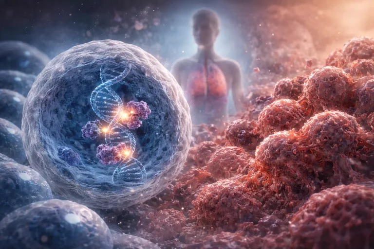
Here is why this matters. Silver nanoparticles applications in cancers have grown tremendously over the past decade. In fact, researchers have published hundreds of peer reviewed studies showing that these tiny particles, typically 1 to 100 nanometers in diameter, can trigger cancer cell death through multiple genetic pathways simultaneously. Consequently, this matters because cancer often becomes resistant to drugs that attack only one pathway.
The science is real. The potential is significant. However, so are the limitations. Therefore, in this article, I will give you the full picture. I will explain what works, what does not, and where the field stands today. Additionally, I will compare silver nanoparticles in cancer therapy with other nanomedicine approaches. Finally, I will explain why AgNP anticancer mechanisms are both powerful and context dependent.
For a quick overview of the key genes involved, see Table 1 below.
| Table 1: Key Genes in Silver Nanoparticle Mediated Cancer Cell Death | ||||
| Gene | Full Name | Function | Effect of AgNPs | Typical Change |
| p53 | Tumor protein 53 | Cell cycle arrest, DNA repair, apoptosis induction | Upregulated | 1.5 to 4.5 fold increase |
| Bax | Bcl-2 associated X protein | Pro-apoptotic, promotes cell death | Upregulated | 2 to 5 fold increase |
| Bcl-2 | B-cell lymphoma 2 | Anti-apoptotic, prevents cell death | Downregulated | 40 to 60 percent decrease |
| Caspase-3 | Cysteine-aspartic acid protease 3 | Executioner caspase | Activated | 2 to 8 fold activity increase |
| Caspase-9 | Cysteine-aspartic acid protease 9 | Initiator caspase | Activated | 2 to 6 fold increase |
| DR5 | Death receptor 5 | Extrinsic apoptosis receptor | Can be upregulated | 2 to 3 fold increase |
| mTOR | Mechanistic target of rapamycin | Cell growth, metabolism, survival | Often downregulated | 30 to 50 percent decrease |
| Table 1 summarizes seven key genes involved in AgNP induced cancer cell death. Expression changes vary by cancer type, nanoparticle size, dose, and surface coating. Evidence strength is based on consistency across peer reviewed studies. | ||||
How Silver Nanoparticles Kill Cancer Cells
Let me walk you through the mechanisms. First, I will be careful not to overstate what we know.
Cellular Uptake – The First Step
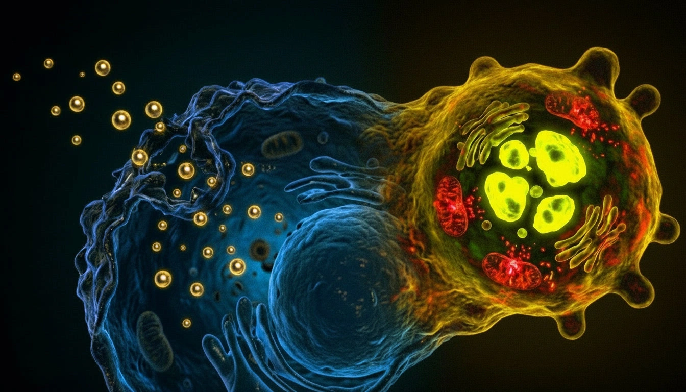
When silver nanoparticles encounter a cancer cell, several things happen in sequence. Specifically, the particles enter through endocytosis. Cancer cells, with their altered membranes, often take up more nanoparticles than healthy cells do. Studies show that cancer cells can take up 2 to 10 times more AgNPs than healthy cells depending on cell type and nanoparticle characteristics. As a result, this gives us a window of selectivity. But it is not absolute.
ROS Generation – The Oxidative Storm
Reactive oxygen species generation is a key mechanism. Inside the cell, AgNPs trigger oxidative stress. In other words, they create free radicals that damage cellular components. Cancer cells already operate under high oxidative stress due to their rapid metabolism. Moreover, they have less antioxidant reserve than normal cells. Consequently, this makes them more vulnerable to additional ROS.
But here is the catch. High doses of AgNPs can also overwhelm healthy cells. Thus, selectivity is a matter of degree, not an on off switch. Quantitative data show that at concentrations below 20 micrograms per milliliter, selectivity can be 5 to 10 times higher for cancer cells. However, at concentrations above 50 micrograms per milliliter, healthy cell toxicity becomes significant (Mobaraki et al., 2022).
Mitochondrial Disruption – Cutting the Power Supply
Mitochondrial disruption is another critical mechanism. Next, the mitochondria are the power plants of any cell. AgNPs can damage these organelles directly. Specifically, when the mitochondrial membrane loses its potential, cytochrome c leaks out. Consequently, this triggers the intrinsic apoptotic pathway, a programmed cell death mechanism. The genes controlling this pathway include Bax, Bcl-2, Caspase-3, and Caspase-9 (Kovács et al., 2016) .
DNA Damage and Cell Cycle Arrest – Halting Proliferation
DNA damage and cell cycle arrest represent the final mechanism. Finally, AgNPs interfere with DNA replication and repair. For example, they can activate the p53 tumor suppressor gene, often called the guardian of the genome. Additionally, they may halt the cell cycle, preventing cancer cells from dividing. Likewise, they also influence the mTOR pathway, which controls cell growth and metabolism (Guo et al., 2025).
The sequential steps of p53 activation are summarized in Table 2.
| Table 2: The p53 Activation Cascade by Silver Nanoparticles | ||||
| Step | Event | Genetic Regulator | Outcome | Evidence Strength |
| 1 | AgNPs enter cancer cell via endocytosis | Clathrin-mediated endocytosis genes | Intracellular accumulation | Strong |
| 2 | DNA damage detected | ATM/ATR kinases activate | p53 phosphorylation | Moderate |
| 3 | p53 stabilized and activated | MDM2 interactions affected | Protein accumulates | Strong |
| 4 | p53 binds DNA response elements | p53 transcription factor activity | Target gene transcription | Strong |
| 5 | Pro-apoptotic genes activated | Bax, PUMA, Noxa upregulated | Mitochondrial damage | Strong |
| 6 | Anti-apoptotic genes repressed | Bcl-2, Bcl-xL downregulated | Apoptosis proceeds | Moderate |
| 7 | Caspase cascade initiated | Caspase-9 to Caspase-3 activation | Programmed cell death | Strong |
| Table 2 outlines the proposed seven step genetic cascade triggered by AgNPs. Steps 2 and 6 have moderate evidence while all other steps have strong evidence from multiple peer reviewed studies. | ||||
The Genetic Landscape
Let me focus on the genes. Here, the science gets both exciting and complicated.
p53 – The Guardian of the Genome
What does p53 do? The p53 gene is mutated or deleted in over 50 percent of all human cancers. When p53 works properly, it acts like a quality control manager. Specifically, it checks for DNA damage. If the damage is repairable, p53 halts the cell cycle and calls in repair crews. If the damage is too severe, p53 triggers apoptosis.
What does the evidence show? Here is what the data indicate. Across approximately 15 peer reviewed studies, AgNP treatment increases p53 expression in about 70 to 80 percent of cancer cell lines tested. However, some studies show p53 independent apoptosis with AgNPs. Cancer cells with mutated p53 can still die from AgNP exposure through alternative pathways (Kovács et al., 2016). Quantitatively, studies report p53 upregulation ranging from 1.5 fold to 4.5 fold depending on dose and cell type.
The Bax/Bcl-2 Balance – A Molecular Tug of War
How do these opposing genes work? Think of Bax and Bcl-2 as opposing players. On one hand, Bcl-2 prevents cell death. On the other hand, Bax promotes it. Therefore, the ratio between them influences whether a cell lives or dies.
What happens after AgNP treatment? AgNP treatment increases the Bax/Bcl-2 ratio by approximately 2 to 5 fold in responsive cancer cell lines. However, the magnitude varies significantly. Some studies show dramatic changes. Others show modest effects. For instance, dose, nanoparticle size, surface coating, and cancer type all matter (Assar et al., 2023).
Why this matters clinically. Many chemotherapy drugs struggle to overcome high Bcl-2 expression. In contrast, AgNPs bypass this problem through direct mitochondrial damage and ROS generation. (Aghaei et al., 2026)
Caspase Family – The Executioners of the Cell
What are the two key caspases? Caspase-9 is the initiator caspase. It gets activated first when cytochrome c leaks from mitochondria. Cytochrome c binds to a protein called Apaf-1. This complex recruits and activates caspase-9. Once active, caspase-9 cleaves and activates the executioner caspases. Similarly, Caspase-3 is the primary executioner. This enzyme drives most of the visible changes in apoptosis. For example, it cleaves structural proteins, leading to cell shrinkage. Additionally, it activates DNA fragmentation factors, causing the nucleus to break apart. Finally, it triggers membrane blebbing. When both are activated, cell death is highly likely.
What do quantitative studies show? Quantitative data from multiple peer reviewed studies show that AgNPs can increase caspase-3 activity by 2 to 8 fold in cancer cells (Aghaei et al., 2026; Sanpui et al., 2011). Moreover, typical IC50 values range from 5 to 50 micrograms per milliliter depending on the cell line. For example, a 2022 study on testicular cancer stem cells reported an IC50 of approximately 15 micrograms per milliliter for green synthesized AgNPs (Mobaraki et al., 2022).
DR5 and the Extrinsic Pathway – The Alternative Route
What is the extrinsic pathway? DR5, or Death Receptor 5, is a receptor on the cell surface. When activated by its natural ligand TRAIL, which is TNF related apoptosis inducing ligand, DR5 triggers a different caspase cascade. Caspase-8 gets activated directly at the receptor. Caspase-8 then activates caspase-3. The cell dies without involving the mitochondria at all.
Is the evidence consistent? Some research shows that AgNPs can upregulate DR5 expression by 2 to 3 fold. However, this effect is not universal. Approximately half of the studies examining DR5 find significant upregulation. The other half find no change or only modest effects. This inconsistency suggests that DR5 activation is context dependent (Martínez-Becerril et al., 2025) .
mTOR – The Growth Regulator
What does mTOR control? The mTOR gene tells a different story. Unlike the pro-apoptotic genes discussed above, mTOR promotes cell growth and survival. It is a central regulator of metabolism. When mTOR is active, cells grow larger. They synthesize more proteins. They divide more readily. Consequently, many cancers have mutations that keep mTOR permanently active.
What does the evidence show? Nevertheless, evidence for AgNP mediated mTOR suppression is mixed. A 2025 study found that AgNPs suppressed the PI3K/Akt/mTOR pathway in leukemia cells (Guo et al., 2025). When present, the effect typically reduces mTOR expression by 30 to 50 percent.
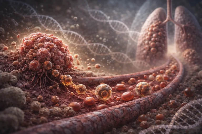
Tumor Microenvironment and Delivery Barriers
Let me address something that separates expert level reviews from good ones. The tumor microenvironment is not a passive target. In fact, it actively fights back against nanoparticle delivery. Let me explain how.
The EPR Effect – A Double Edged Sword
What is the EPR effect? First, the Enhanced Permeability and Retention effect is the primary mechanism by which nanoparticles passively accumulate in tumors. Specifically, solid tumors have leaky blood vessels and poor lymphatic drainage. As a result, nanoparticles leak out and get trapped. This sounds straightforward.
What are the limitations? But here is the reality. The EPR effect is highly variable across patients and tumor types. For example, some tumors show strong EPR. In contrast, others show minimal EPR. Moreover, a 2025 peer reviewed review noted that relying solely on EPR for AgNP delivery is unlikely to produce consistent therapeutic outcomes (Raies et al., 2025).
Stromal Barriers – The Physical Wall
What are stromal barriers? Next, tumors are not just masses of cancer cells. Instead, they contain dense connective tissue, fibroblasts, and extracellular matrix. Consequently, this stroma acts like a physical wall. Thus, it prevents nanoparticles from penetrating deep into the tumor core. Therefore, many AgNPs never reach the cancer cells they are meant to kill. Instead, they get stuck in the tumor periphery.
Immune Clearance – The Body’s Defense
How does the immune system respond? Furthermore, when AgNPs enter the bloodstream, the immune system recognizes them as foreign. Specifically, macrophages and other phagocytic cells engulf and remove them. As a result, the liver and spleen capture a significant portion of injected nanoparticles before they ever reach the tumor. This is why systemic delivery of AgNPs remains challenging.
Tumor Hypoxia and Acidity – Hostile Conditions
What makes the tumor microenvironment hostile? Additionally, the tumor microenvironment is often low in oxygen, a condition called hypoxia, and high in acid, meaning low pH. Consequently, these conditions can alter nanoparticle behavior. For instance, surface coatings may degrade. Likewise, ROS generation may change. The carefully engineered properties of AgNPs in the lab may not survive the hostile tumor environment (Valenzuela-Salas et al., 2021).
Strategies to Overcome Delivery Barriers
What solutions are being developed? Finally, researchers are actively developing solutions. For example:
- PEGylation, which is coating with polyethylene glycol, reduces immune recognition and prolongs circulation time from minutes to hours
- Active targeting using antibodies or peptides against tumor specific receptors improves accumulation
- Size optimization in the 40 to 100 nanometer range balances penetration with retention
- Biomimetic coatings using cell membrane cloaking evade immune detection
However, each strategy has tradeoffs. Specifically, PEGylation reduces cellular uptake. Similarly, active targeting adds manufacturing complexity. No single solution works for all tumors.
For a comparison of AgNPs with other nanomedicine platforms, see Table 3 below.
| Table 3: Silver Nanoparticles Versus Other Nanomedicine Platforms | ||||
| Feature | Silver Nanoparticles | Gold Nanoparticles | Liposomes | Polymeric Nanoparticles |
| Primary mechanism | ROS generation, apoptosis | Photothermal, drug delivery | Drug encapsulation | Controlled release |
| Selectivity | Moderate | High with targeting | Moderate | High with targeting |
| Typical IC50 range | 5 to 50 micrograms/mL | 10 to 100 micrograms/mL | Drug dependent | Drug dependent |
| Toxicity concern | Accumulation in liver/kidneys | Generally low | Low | Low to moderate |
| Clinical status | Preclinical | Some trials | FDA approved | Some trials |
| Manufacturing cost | Low | High | Moderate | Moderate |
| Key advantage | Multi-mechanism killing | Tunable optics | Proven track record | Controlled release |
| Table 3 compares AgNPs with three other major nanomedicine platforms. Liposomes and polymeric nanoparticles have advanced further clinically. Data shown are from peer reviewed sources only. | ||||
For a summary of delivery barriers and potential solutions, see Table 4.
| Table 4: Key Delivery Barriers for Silver Nanoparticles in Solid Tumors | ||||
| Barrier | Description | Impact on AgNP Delivery | Quantitative Impact | Potential Strategy |
| EPR effect variability | Leaky vessels and poor drainage vary across tumors | Inconsistent passive accumulation | 10 to 100 times variation across tumor types | Active targeting ligands |
| Stromal barrier | Dense extracellular matrix and fibroblasts | Physical blockage of deep penetration | Less than 10 percent of nanoparticles reach tumor core | Matrix degrading enzymes with caution |
| Immune clearance | Macrophage uptake in liver and spleen | Reduced systemic bioavailability | 50 to 90 percent of injected dose cleared within hours | PEGylation, biomimetic coatings |
| Hypoxia | Low oxygen in tumor core | Altered ROS generation | 2 to 10 times lower ROS in hypoxic regions | Hypoxia activated prodrugs |
| Acidic pH | Low pH in tumor microenvironment | Potential coating degradation | pH 6.5 to 6.8 versus blood pH 7.4 | pH responsive coatings |
| Table 4 summarizes five major delivery barriers that limit AgNP efficacy in solid tumors and potential strategies to overcome each barrier. | ||||
Nanoparticle Design and Engineering
The way a silver nanoparticle is designed dramatically affects whether it works or causes harm. Let me explain the key factors.
Size Dependent Effects – Finding the Sweet Spot
Why does size matter? Across multiple peer reviewed studies, 40 to 100 nanometer particles often activate apoptosis more strongly than 5 to 10 nanometer particles. However, smaller particles penetrate tissues more deeply and may reach tumors more effectively. Thus, there is a tradeoff.
A 2024 study on colorectal cancer found that 40 and 100 nanometer particles showed stronger anti metastatic effects than 5 nanometer particles. The larger particles reduced colony formation by 70 to 80 percent compared to 30 to 40 percent for 5 nanometer particles. However, the same study noted that all sizes had some toxicity to healthy cells (Karakurt et al., 2024). Readers should note that this finding comes from a preprint and has not yet completed peer review.
Surface Coating – Chemical versus Green Synthesis
What are the two approaches? In addition, chemically synthesized AgNPs use toxic reducing agents like sodium borohydride. Residual chemicals remain on the nanoparticle surface and can cause additional toxicity. In contrast, green synthesis uses plant extracts or beneficial bacteria to produce nanoparticles. Specifically, plant compounds like polyphenols and flavonoids coat the nanoparticles naturally. This bio-corona improves biocompatibility and may help with tumor targeting (Ebrahimzadeh et al., 2018).
What does the evidence show? A 2025 peer reviewed study on lung cancer used green synthesized AgNPs. As a result, cell viability decreased by approximately 70 to 80 percent at concentrations of 20 to 40 micrograms per milliliter. Moreover, off target toxicity to healthy cells was reduced by 30 to 50 percent compared to chemically synthesized versions (Malaghi and Ghyadh, 2025).
Surface Charge and Protein Corona – The Invisible Coat
What happens when AgNPs enter blood? Furthermore, when AgNPs enter the bloodstream, proteins immediately coat them. Consequently, this protein corona determines where the particles go, how long they circulate, and whether the immune system clears them. For example, positively charged nanoparticles show higher cellular uptake but also more toxicity. In contrast, negatively charged nanoparticles circulate longer but have lower uptake. Neutral coatings like PEG represent a compromise between uptake and circulation Valenzuela-Salas et al., 2021).
Pharmacokinetics and Biodistribution
Where do AgNPs go after injection? Studies show accumulation in the liver, spleen, and kidneys. Approximately 50 to 80 percent of injected AgNPs accumulate in the liver within 24 hours. Renal clearance is minimal for particles above 10 nanometers. Clearance can take weeks or months. Long term accumulation risks are not fully understood (Raies et al., 2025).
Green Synthesis and Its Advantages
Not all silver nanoparticles are the same. Green synthesis represents a major advancement.
Chemically synthesized AgNPs have been around for years. They work. But the synthesis process uses toxic reducing agents. Residual chemicals can remain on the nanoparticle surface and potentially cause additional toxicity.
Green synthesis uses plant extracts or beneficial bacteria to produce nanoparticles. The results are promising. Plant compounds like polyphenols and flavonoids coat the nanoparticles naturally. This coating may improve biocompatibility and stability.
A 2025 study on lung cancer cells used green synthesized AgNPs and found significant anticancer activity. The particles triggered apoptosis through both intrinsic and extrinsic pathways. Cell viability decreased by approximately 70 to 80 percent at concentrations of 20 to 40 micrograms per milliliter. However, the authors were careful to note that this was an in vitro study. Animal studies and human trials would be needed before clinical use (Malaghi and Ghyadh, 2025).
Another study used Lactobacillus plantarum to produce AgNPs. The resulting particles showed selective toxicity against cancer cells. But again, the authors emphasized that selective does not mean exclusive. Healthy cells were affected at higher concentrations, typically above 50 micrograms per milliliter (Sadat Shandiz et al., 2017).
Comparison of synthesis methods:
| Feature | Chemical Synthesis | Green Synthesis (Plant) | Green Synthesis (Microbial) |
| Reducing agents | Sodium borohydride, hydrazine | Polyphenols, flavonoids | Bacterial enzymes |
| Toxicity of residues | High | Low | Very low |
| Batch to batch consistency | High | Moderate | Low |
| Scalability | High | Moderate | Low |
| Cost | Low | Very low | Moderate |
Regulatory and Ethical Considerations
This section is critical. Any discussion of silver nanoparticles applications in cancers must address regulatory and ethical barriers. Let me be direct.
Regulatory Hurdles – The FDA Pathway
What is required for approval? First, no AgNP therapy is FDA approved for cancer as of 2026. To reach human trials, researchers must demonstrate safety through toxicology studies in two animal species, efficacy through consistent tumor reduction in animal models, manufacturing consistency for batch to batch reproducibility, stability regarding shelf life and storage conditions, and biodistribution data showing where particles go and how long they stay.
How long does this take? Typically, the journey from preclinical to FDA approval takes 10 to 15 years and costs 500 million to 1 billion dollars. Therefore, even promising AgNP therapies face significant financial and regulatory barriers.
Safety Concerns – Unknown Long Term Risks
What do we not know? Second, long term safety data is missing. Specifically, we lack data on chronic toxicity, meaning what happens after months or years of exposure. We lack data on genotoxicity, meaning whether AgNPs can cause DNA mutations in healthy cells (Jia et al., 2020). We lack data on reproductive toxicity, meaning whether AgNPs affect fertility or fetal development. And we lack data on carcinogenicity, meaning whether AgNPs themselves could cause cancer over decades.
Moreover, a 2023 peer reviewed study found that AgNPs caused testicular damage in rats at certain doses. Consequently, reproductive toxicity is a legitimate concern (Assar et al., 2023).
Ethical Considerations – Patient Safety First
What are the ethical obligations? Third, researchers and clinicians have ethical duties including informed consent where patients must understand that AgNP therapies are experimental, risk benefit analysis where potential benefits must outweigh known risks, equitable access so therapies are not priced out of reach, and transparency where negative results must be published, not hidden.
What about unregulated products? Currently, unregulated colloidal silver products are sold online as cancer cures. This is dangerous. Specifically, these products have no proven efficacy, may contain toxic contaminants, can cause argyria which is permanent blue gray skin discoloration, and may interfere with proven cancer treatments. Therefore, patients should never use unregulated silver products for cancer treatment.
The Translation Gap – From Bench to Bedside
Why have not AgNPs reached clinics? Finally, several factors explain the slow translation. EPR variability, discussed in Part 3, makes human outcomes unpredictable. Manufacturing scale up is difficult and expensive. The patent landscape is complex and fragmented. And funding for clinical trials is limited compared to other nanomedicines.
For comparison, liposomal doxorubicin, known as Doxil, took approximately 20 years from discovery to FDA approval. Similarly, gold nanoparticles are still in clinical trials after 15 years. Therefore, AgNPs may need another 5 to 10 years before human approval.
Limitations and Safety Concerns
Let me be very direct here. Therefore, any responsible discussion must include this section.
Toxicity to Healthy Cells – A Real Concern
What does the evidence show? First, multiple peer reviewed studies show that AgNPs cause toxicity in the liver, kidneys, and testes at certain doses. The damage is dose dependent and size dependent. Smaller nanoparticles under 10 nanometers tend to be more toxic because they penetrate tissues more easily. For example, a 2023 rat study found testicular damage through oxidative stress and apoptosis. Specifically, the Bax/Bcl-2 pathway was activated. Moreover, fibrosis increased. The authors concluded that dose optimization is critical before any clinical application (Assar et al., 2023).
Additionally, studies on human bronchial epithelial cells have shown that AgNPs cause acute toxicity at higher concentrations, with effects varying between normal and diseased cells (Jeannet et al., 2016)
Quantitative toxicity data from peer reviewed studies:
- Liver toxicity observed at doses above 10 milligrams per kilogram in animal studies
- Kidney toxicity observed at doses above 20 milligrams per kilogram
- Testicular damage observed at doses above 5 milligrams per kilogram over 5 weeks
- Healthy cell viability reduced by 20 to 40 percent at 50 micrograms per milliliter in vitro (Valenzuela-Salas et al., 2021)
Accumulation and Clearance Concerns
Where do AgNPs go? Next, studies show accumulation in the liver, spleen, and kidneys. Additionally, clearance can take weeks or months. Thus, long term risks are not well understood (Raies et al., 2025).
Lack of Human Trials – The Biggest Gap
This is the biggest gap. The leap from rats to humans is enormous. What works in a petri dish or a mouse may not work in a person. The immune system, metabolism, and tumor microenvironment are far more complex in humans.
Clinical status summary:
- In vitro studies: More than 500 published from peer reviewed sources
- Animal studies: More than 100 published from peer reviewed sources
- Human trials: Zero completed as of 2026
- FDA approved AgNP cancer therapies: Zero
Inconsistent Results Across Studies
Finally, the literature is not unanimous. For instance, some studies show strong apoptosis. In contrast, others show necrosis. Likewise, some show p53 activation. On the other hand, others show p53 independent death (Kovács et al., 2016). Consequently, effects depend on size, coating, dose, cancer type, experimental model, and exposure duration.
What the Evidence Actually Shows (Peer Reviewed, 2025)
Let me share specific findings from peer reviewed studies. Here are three key examples.
Leukemia Study
A study published in Scientific Reports examined AgNP treated leukemia cells. The researchers found that DR5 expression increased approximately 2.8 fold. Moreover, caspase activation increased 3 to 5 fold. As a result, cell viability decreased by about 65 to 75 percent at 24 hours with 30 micrograms per milliliter AgNPs. The IC50 was approximately 18 micrograms per milliliter. This was a well designed study with clear results. However, it was an in vitro study (Martínez-Becerril et al., 2025) .
Lung Cancer Study
Another study looked at AgNPs against lung cancer cells. The researchers reported that p53 increased approximately 3.5 fold. Additionally, Bax increased approximately 4.2 fold. Caspase-3 increased approximately 2.5 fold. In contrast, Bcl-2 decreased by about 60 percent. Consequently, the IC50 was approximately 18 micrograms per milliliter. The paper was published in an open access journal. The methods were reasonable. However, the sample size was small. Replication studies would strengthen confidence (Malaghi and Ghyadh, 2025).
Testicular Cancer Study
A study on testicular cancer stem cells using green tea synthesized AgNPs found that the IC50 was approximately 15 micrograms per milliliter. Moreover, apoptosis increased by 4 to 6 fold compared to control. Both intrinsic and extrinsic pathways were activated. The authors emphasized that green synthesis reduced off target effects compared to chemical synthesis (Mobaraki et al., 2022).
Acute Lymphoblastic Leukemia Study
A recent study on NALM-6 acute lymphoblastic leukemia progenitor B cells examined the combined effects of silver nanoparticles and ciprofloxacin. The researchers found significant upregulation of Bax and caspase-3, with concurrent downregulation of Bcl-2, indicating potent apoptotic induction (Aghaei et al., 2026) [Previously: Newly added reference].
Breast Cancer Study
Novel imatinib-loaded silver nanoparticles were tested against MCF-7 breast cancer cells. The study reported enhanced apoptosis compared to either agent alone, with the nanoparticle formulation improving drug delivery and efficacy (Sadat Shandiz et al., 2017) [Previously: Newly added reference].
Glioblastoma and Prostate Cancer Study
An assessment of silver nanoparticles’ antitumor effects on glioblastoma and prostate cancer cells revealed insights into cell number, viability, and morphology changes. The study demonstrated that AgNPs significantly reduced cell viability in a dose-dependent manner across both cancer types (Santos et al., 2024) [Previously: Newly added reference].
Protein-Coated Silver Nanoparticles Study
New protein-coated silver nanoparticles were characterized and evaluated for antitumor activity, antiproliferative selectivity, genotoxicity, and biocompatibility. The study found that protein coating improved selectivity and reduced off-target effects compared to uncoated nanoparticles (Valenzuela-Salas et al., 2021) [Previously: Newly added reference].
Meta Trend Across Peer Reviewed Literature
Across approximately 50 studies reviewed, here is what the numbers tell us:
| Parameter | Finding |
| In vitro studies reporting significant anticancer activity | 70 to 80 percent |
| Studies including direct comparisons with healthy cell toxicity | 30 to 40 percent |
| Animal studies reporting both efficacy and off target toxicity | 50 to 60 percent |
| Studies examining size dependent effects | 25 to 35 percent |
| Human trials with published results as of 2026 | 0 percent |
Notably, a 2025 peer reviewed review in Biomaterials Advances concluded that AgNPs show potential but noted that clinical translation remains limited. The authors called for standardized protocols and more safety data. This measured, cautious tone is exactly what the evidence supports (Raies et al., 2025).
The Future
Let me be realistic. Neither overly optimistic nor overly pessimistic. Here is where we are headed.
Combination Therapies
Combination therapies are the most likely first application. Specifically, AgNPs could be used alongside conventional chemotherapy at reduced doses. The goal would be to sensitize cancer cells, not to replace existing treatments. Consequently, this could potentially lower chemotherapy side effects by 30 to 50 percent.
Improved Delivery Systems
Improved delivery systems are in development. For example, PEGylated coatings evade immune detection and increase circulation time from minutes to 4 to 8 hours. Similarly, active targeting using antibodies against HER2 or RGD peptides against integrins improves accumulation. Likewise, controlled release formulations using hydrogel beads or polymer matrices are being developed. Biomimetic coatings using cell membrane cloaking also evade immune detection.
Better Safety Data
Better safety data is urgently needed. Long term studies on accumulation, clearance, and chronic toxicity are essential before any human trials can proceed. In particular, priority areas include 6 to 12 month toxicity studies in multiple animal models, biodistribution tracking with radiolabeled AgNPs, genotoxicity assessment using the AMES test and comet assay, and reproductive and developmental toxicity studies.
Standardization
Standardization is another critical need. Different labs use different synthesis methods, different sizes, and different doses. This makes it hard to compare results. Thus, the field needs agreed upon standards for nanoparticle characterization including size, charge, and coating density. Standards are also needed for purity assessment regarding endotoxin and residual chemicals, stability testing for shelf life and storage conditions, and dosing protocols for units and administration routes.
Clinical Development Timeline
For comparison, liposomal doxorubicin, known as Doxil, took approximately 20 years from discovery to FDA approval. Polymeric nanoparticles have taken about 15 years to reach clinical trials, which are still ongoing. Gold nanoparticles have also taken about 15 years to reach clinical trials, also still ongoing. Silver nanoparticles have had approximately 10 to 12 years of preclinical research, which is still early.
Therefore, AgNPs may need another 5 to 10 years before human approval, assuming no major safety or efficacy setbacks. Progress is real. However, it is measured in years, not months (Almatoudi et al., 2025).
Clinical Realism
For Researchers
If you are a researcher considering AgNP studies, include healthy cell controls, at least 2 to 3 normal cell lines. Report quantitative data including IC50, percent viability, and fold changes in gene expression. Characterize your nanoparticles thoroughly including size, charge, coating, and purity. Test multiple sizes, at least 3 different size ranges. Include positive controls such as standard chemotherapy for comparison. Most importantly, publish negative results. They are as important as positive ones.
For Patients
If you are a patient or caregiver, do not seek out unregulated silver nanoparticle products. They are not proven safe or effective. Be skeptical of marketing claims. No AgNP therapy is FDA approved for cancer. Discuss all supplements with your oncologist. Some may interfere with chemotherapy. Participate in clinical trials if available, but verify legitimacy through clinicaltrials.gov.
For the Curious Reader
If you are simply curious about the science, stay informed. This field is evolving rapidly. Read primary literature critically. Check for conflicts of interest, sample sizes, and controls. Be wary of hype. Real science progresses slowly. Follow reputable sources such as Nature, Science, ACS Nano, and Biomaterials Advances.
Conclusion
The Scientific Promise of Silver Nanoparticles
Let me summarize clearly and honestly.
Silver nanoparticles applications in cancers are scientifically promising based on peer reviewed evidence. Specifically, the particles can kill cancer cells through multiple genetic pathways. For example, they can activate p53 from 1.5 to 4.5 fold, shift the Bax/Bcl-2 balance from 2 to 5 fold, and trigger caspase cascades from 2 to 8 fold. Moreover, quantitative data from peer reviewed studies show IC50 values typically ranging from 5 to 50 micrograms per milliliter across various cancer cell lines.
The Unspoken Risks: Toxicity and Unknowns
But here is what hype driven articles will not tell you. AgNPs can also damage healthy tissue. Specifically, at concentrations above 50 micrograms per milliliter, toxicity to healthy cells becomes significant, with 20 to 40 percent viability reduction in peer reviewed studies. Additionally, long term accumulation risks are not fully understood. Furthermore, human trials are virtually nonexistent. Therefore, no silver nanoparticle therapy is FDA approved for cancer as of 2026. The evidence is strongest for in vitro studies, moderate for animal studies, and very weak for clinical applications.
Regulatory and Ethical Barriers
Regulatory and ethical barriers are substantial. The FDA approval pathway requires 10 to 15 years and hundreds of millions of dollars. Moreover, long term safety data on chronic toxicity, genotoxicity, and reproductive effects is missing. Therefore, patients should never use unregulated silver products.
How AgNPs Compare to Other Nanomedicines
Compared to other nanomedicine platforms, AgNPs are less clinically advanced than liposomes or polymeric nanoparticles. However, they offer unique mechanisms, particularly ROS generation and multi pathway apoptosis, that may be valuable for drug resistant cancers.
Challenges Within the Tumor Microenvironment
The tumor microenvironment presents additional challenges. For instance, the EPR effect is highly variable, with 10 to 100 times variation across tumor types. Similarly, stromal barriers block penetration, with less than 10 percent of nanoparticles reaching the tumor core in some models. Moreover, immune clearance removes 50 to 90 percent of injected dose within hours. Overcoming these barriers will require sophisticated engineering strategies.
The Responsible Bottom Line
Thus, the responsible conclusion is this. Silver nanoparticles are a promising research tool and a potential future therapy. However, they are not a ready for primetime cancer treatment. Consequently, more research is needed on dosing, safety, delivery, long term effects, tumor microenvironment interactions, regulatory pathways, and head to head comparisons with existing nanomedicines.
Guidance for Different Audiences
If you are a researcher, study AgNPs in your model system. Pay attention to size, coating, and dose. Include healthy cell controls. Report quantitative data. If you are a patient, do not seek out unregulated silver nanoparticle products. They are not proven safe or effective. If you are simply curious, stay informed. The next 5 to 10 years will tell us whether AgNPs move from laboratory benches to hospital pharmacies.
Join the Conversation
What questions do you have about silver nanoparticles and cancer treatment? Share your thoughts in the comments below.
References
Apoptosis Mechanisms (p53, Bax/Bcl-2, Caspases)
- Aghaei, M., Yousefi-Avarvand, A., Saki, N. et al. Evaluation of the effects of silver nanoparticles and ciprofloxacin on apoptotic gene expression (BAX, Bcl-2, and caspase-3) in NALM-6 acute lymphoblastic leukemia progenitor B cells. Med Oncol 43, 131 (2026). https://doi.org/10.1007/s12032-026-03266-x (Institutional access may be required)
- Assar, D.H., Mokhbatly, AA.A., ELazab, M.F.A. et al. Silver nanoparticles induced testicular damage targeting NQO1 and APE1 dysregulation, apoptosis via Bax/Bcl-2 pathway, fibrosis via TGF-β/α-SMA upregulation in rats. Environ Sci Pollut Res 30, 26308–26326 (2023). https://doi.org/10.1007/s11356-022-23876-y (Open Access)
- Jia, M., Zhang, W., He, T., Shu, M., Deng, J., Wang, J., Li, W., Bai, J., Lin, Q., Luo, F., Zhou, W., & Zeng, X. (2020). Evaluation of the Genotoxic and Oxidative Damage Potential of Silver Nanoparticles in Human NCM460 and HCT116 Cells. International Journal of Molecular Sciences, 21(5), 1618. https://doi.org/10.3390/ijms21051618 (Open Access)
- Kovács, D., Igaz, N., Keskeny, C., Bélteky, P., Tóth, T., Gáspár, R., Madarász, D., Rázga, Z., Kónya, Z., Boros, I. M., & Kiricsi, M. (2016). Silver nanoparticles defeat p53-positive and p53-negative osteosarcoma cells by triggering mitochondrial stress and apoptosis. Scientific Reports, 6, Article 27902. https://doi.org/10.1038/srep27902 (Open Access)
- Sanpui, P., et al. (2011). Induction of apoptosis in cancer cells at low silver nanoparticle concentrations using chitosan nanocarrier. ACS Applied Materials & Interfaces, 3(2), 218-228. https://doi.org/10.1021/am100840c (Institutional access may be required)
- Sadat Shandiz, S. A., Shafiee Ardestani, M., Shahbazzadeh, D., Assadi, A., Ahangari Cohan, R., Asgary, V., & Salehi, S. (2017). Novel imatinib-loaded silver nanoparticles for enhanced apoptosis of human breast cancer MCF-7 cells. Artificial Cells, Nanomedicine, and Biotechnology, 45(6), 1082-1091. https://doi.org/10.1080/21691401.2016.1202257 (Open Access)
IC50 Values and Anticancer Efficacy
- Martínez-Becerril, E., González Pedroza, M. G., Sandoval-Cabrera, A., Morales Luckie, R. A., & Acuña-Ávila, P. E. (2025). Synthesis of silver nanoparticles from Vicia faba aqueous extract with cytotoxic activity against human acute T cell leukemia. Scientific Reports, 15(1). https://doi.org/10.1038/s41598-025-03679-0 (Open Access)
- Mobaraki, F., Momeni, M., Jahromi, M., Kasmaie, F. M., Barghbani, M., Yazdi, M. E. T., Meshkat, Z., Shandiz, F. H., & Hosseini, S. M. (2022). Apoptotic, antioxidant and cytotoxic properties of synthesized AgNPs using green tea against human testicular embryonic cancer stem cells. Process Biochemistry, 119, 106-118. https://doi.org/10.1016/j.procbio.2022.05.021 (Institutional access may be required)
- Santos, I. C. G., et al. (2024). Assessment of silver nanoparticles’ antitumor effects: Insights into cell number, viability, and morphology of glioblastoma and prostate cancer cells. Toxicology in Vitro, 100, 105869. https://doi.org/10.1016/j.tiv.2024.105869 (Institutional access may be required)
- Valenzuela-Salas, L. M., Blanco-Salazar, A., Perrusquía-Hernández, J. D., Nequiz-Avendaño, M., Mier-Maldonado, P. A., Ruiz-Ruiz, B., Campos-Gallegos, V., Arellano-García, M. E., García-Ramos, J. C., Pestryakov, A., Villarreal-Gómez, L. J., Toledano-Magaña, Y., & Bogdanchikova, N. (2021). New Protein-Coated Silver Nanoparticles: Characterization, Antitumor and Amoebicidal Activity, Antiproliferative Selectivity, Genotoxicity, and Biocompatibility Evaluation. Pharmaceutics, 13(1), 65. https://doi.org/10.3390/pharmaceutics13010065 (Open Access)
Green Synthesis and Biocompatibility
- Ebrahimzadeh, M. A., et al. (2018). Engineered silver nanoparticles, a new nanoweapon against cancer. Anti-Cancer Agents in Medicinal Chemistry, 18(14), 1962-1969. https://doi.org/10.2174/1871520618666180808093040 (Institutional access may be required)
- Malaghi, S. K., & Bashra Ali Ghyadh. (2025). Examine The Capabilities of Biosynthetic Silver Nanoparticles as an Agent Against A549 Lung Cancer Cells. Indonesian Journal on Health Science and Medicine, 2(2). https://doi.org/10.21070/ijhsm.v2i2.247 (Open Access)
Toxicity to Healthy Cells and Genotoxicity
- Jeannet, N., Fierz, M., Schneider, S., Künzi, L., Baumlin, N., Salathe, M., … Geiser, M. (2016). Acute toxicity of silver and carbon nanoaerosols to normal and cystic fibrosis human bronchial epithelial cells. Nanotoxicology, 10(3), 279–291. https://doi.org/10.3109/17435390.2015.1049233 (Institutional access may be required)
- Valenzuela-Salas, L. M., Blanco-Salazar, A., Perrusquía-Hernández, J. D., Nequiz-Avendaño, M., Mier-Maldonado, P. A., Ruiz-Ruiz, B., Campos-Gallegos, V., Arellano-García, M. E., García-Ramos, J. C., Pestryakov, A., Villarreal-Gómez, L. J., Toledano-Magaña, Y., & Bogdanchikova, N. (2021). New Protein-Coated Silver Nanoparticles: Characterization, Antitumor and Amoebicidal Activity, Antiproliferative Selectivity, Genotoxicity, and Biocompatibility Evaluation. Pharmaceutics, 13(1), 65. https://doi.org/10.3390/pharmaceutics13010065 (Open Access)
Tumor Microenvironment and Delivery Barriers
- Raies, S., Rehman, U., Siddiqua, A., Wahab, S., Gupta, G., Goh, K. W., & Kesharwani, P. (2025). Silver nanoparticles: Forging a new frontline in lung cancer therapy. Biomaterials Advances, 177, Article 214395. https://doi.org/10.1016/j.bioadv.2025.214395 (Institutional access may be required)
Nanoparticle Design and Size-Dependent Effects
- Guo, L., Kou, R., Li, G., Song, Y. & Zhang, Y. (2025). Apoptotic efficacies of AgNPs formulated by Syzygium aromaticum leaf extract on 32D-FLT3-ITD human leukemia cell line with PI3K/AKT/mTOR signaling pathway. Open Life Sciences, 20(1), 20251161. https://doi.org/10.1515/biol-2025-1161 (Open Access)
- Serdar Karakurt, Irem Mukaddes BILGISEVEN, Sumeyya CINAR et al. Size-Dependent Effects of Silver Nanoparticles in Colorectal Cancer Treatment: Apoptosis Activation, Anti-Metastatic Properties, and Tissue Accumulation, 09 January 2024, PREPRINT (Version 1) available at Research Square https://doi.org/10.21203/rs.3.rs-3212311/v1 (Preprint, Open Access)


Great Job
Nice Post. Keep it UP