
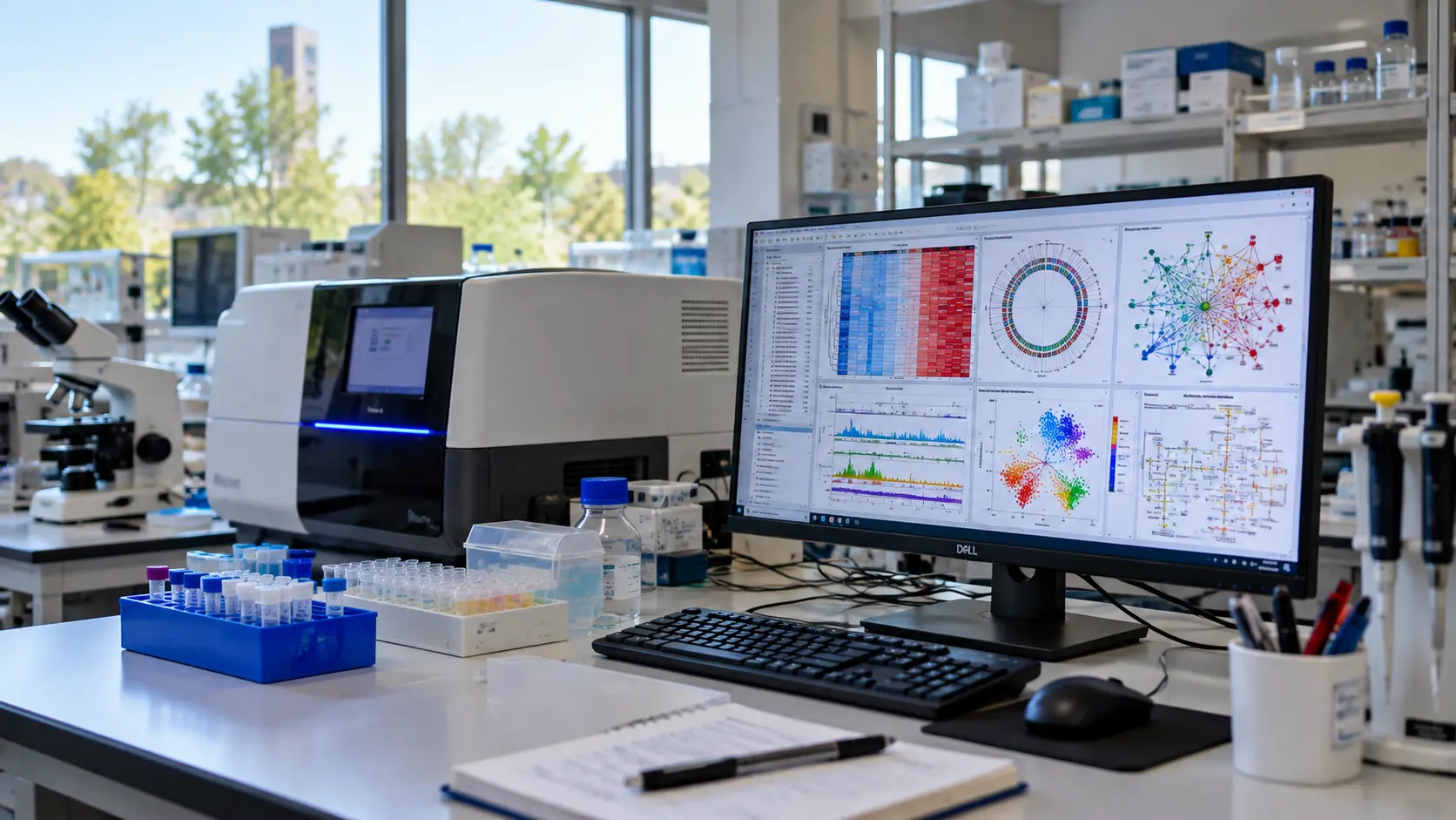
Omics Technologies: Exploring Biology at Multiple Molecular Levels
Introduction
What if scientists could investigate the DNA, RNA, proteins, and metabolites of a biological system to understand how its components work together? This is the central idea behind Omics Technologies, a collection of high-throughput methods that measure large sets of biological molecules. Instead of studying one gene or protein at a time, researchers can examine multiple molecular features simultaneously.
This approach matters because biological processes rarely depend on a single molecule. Cancer, cardiovascular disorders, diabetes, and inherited diseases involve interactions among genetic variants, gene expression, protein activity, metabolism, and environmental influences. Studying only one molecular layer may therefore provide an incomplete explanation.
Different omics approaches answer different questions. Genomics examines DNA sequence and genetic variation. Transcriptomics investigates RNA expression. Proteomics measures proteins. Metabolomics characterizes small molecules involved in cellular metabolism. Epigenomics, metagenomics, lipidomics, and other specialized fields provide additional perspectives (Gottardi Zamperla et al., 2026).
Recent advances in single-cell sequencing, spatial molecular profiling, mass spectrometry, and artificial intelligence have expanded what Omics Technologies can achieve. However, these methods also present challenges involving data quality, experimental design, computational complexity, cost, and clinical validation.
This guide explains the major omics technologies, how they work, where they are applied, and how researchers combine them to investigate biological questions.
What Are Omics Technologies?
Omics Technologies are high-throughput approaches used to characterize large sets of biological molecules, including DNA, RNA, proteins, lipids, metabolites, and epigenetic features. The suffix -omics generally refers to the comprehensive study of a particular class of biological components, while -ome describes the corresponding collection, such as the genome or proteome.
Each omics discipline captures a different aspect of biological organization. Because these measurements reflect different biological processes, no single omics layer provides a complete explanation of a complex biological system.
The Core Omics Layers
Genomics
Genomics studies the genome, including DNA sequence, genetic variants, and structural changes. Whole-genome sequencing (WGS) examines most of the genome, whereas whole-exome sequencing (WES) focuses on protein-coding regions. These methods help identify disease-associated variants, investigate inherited disorders, and characterize cancer genomes (Gottardi Zamperla et al., 2026).
Transcriptomics
Transcriptomics examines RNA molecules and helps researchers understand which genes are expressed under particular conditions. RNA sequencing can characterize gene-expression patterns, alternative splicing, and changes associated with disease. Single-cell RNA sequencing resolves expression differences among individual cells, while spatial transcriptomics adds information about the locations of RNA molecules within tissue.
Proteomics
Proteomics investigates the identity, abundance, and characteristics of proteins. Mass spectrometry is widely used to identify and quantify thousands of proteins, while antibody-based techniques can measure selected proteins or map their distribution within tissues. Proteomics can also investigate post-translational modifications, including phosphorylation and glycosylation (Dong et al., 2026).
Epigenomics
Epigenomics examines molecular features associated with the regulation of genome activity without requiring changes to the underlying DNA sequence. These features include DNA methylation, histone modifications, and chromatin accessibility. Bisulfite sequencing and related methods can assess DNA methylation. ChIP-seq and CUT&Tag can profile selected histone modifications or DNA-associated proteins. ATAC-seq measures chromatin accessibility. These methods provide complementary information about gene regulation and cellular identity.
Metabolomics
Metabolomics characterizes small molecules, such as sugars, amino acids, organic acids, and other metabolites, in biological samples. Liquid chromatography–mass spectrometry (LC-MS) and nuclear magnetic resonance (NMR) spectroscopy are commonly used approaches. Metabolomic profiles can reveal biochemical changes associated with disease, nutrition, drug exposure, or environmental conditions. Because metabolite levels change with sampling conditions and physiological state, careful sample collection and standardized analysis are essential.
Together, these core disciplines provide complementary views of biological systems. Genomics identifies potential genetic influences. Transcriptomics examines RNA expression. Proteomics investigates proteins. Epigenomics explores regulatory features. Metabolomics characterizes biochemical states (Yiu et al., 2025).
Beyond the Core Omics Layers
The omics landscape extends beyond these five core disciplines. Several specialized fields address biological questions that cannot be answered adequately by core approaches alone.
Metagenomics
Metagenomics studies genetic material recovered directly from microbial communities, including organisms that are difficult to culture in the laboratory. Shotgun metagenomic sequencing can characterize microbial diversity and identify genes associated with metabolic pathways, antimicrobial resistance, and other biological functions. Amplicon sequencing, such as 16S rRNA gene profiling, provides a more targeted approach (Baião et al., 2025).
Lipidomics
Lipidomics investigates the composition, abundance, and biological roles of lipids. These molecules are essential components of cell membranes and participate in energy storage, signaling, inflammation, and cellular communication. Lipidomics is particularly relevant to metabolic disorders, cardiovascular disease, and cancer biology (Kim et al., 2026).
Glycomics
Glycomics examines glycans, the carbohydrate structures attached to proteins and lipids. Glycan composition influences protein stability, cellular recognition, immune responses, and host–pathogen interactions. Glycomics is therefore important in immunology and biopharmaceutical research (Dong et al., 2026).
Interactomics
Interactomics investigates molecular interactions, including protein–protein interactions and relationships among proteins, nucleic acids, and other cellular components. Techniques such as affinity-purification mass spectrometry help researchers identify molecular networks involved in signaling and disease.
Phenomics
Phenomics studies biological traits on a broad scale, often using imaging, physiological measurements, and computational analysis. When combined with genomic information, phenomics can help connect genetic variation with observable characteristics.
These disciplines overlap rather than forming independent categories. The appropriate combination depends on the biological question, sample type, and available methods.
The following table 1 provides a structured summary of the major omics technologies, the biological features they investigate, their representative methods, and their example applications, thereby consolidating the core and specialized disciplines discussed above.
| Table 1. Major Omics Technologies, Methods, and Applications | |||
| Omics discipline | What it investigates | Representative methods | Example applications |
| Genomics | DNA sequence and genetic variation | Whole-genome sequencing, WES | Inherited disorders, cancer variants, population genetics |
| Transcriptomics | RNA abundance and transcript structure | RNA-seq, single-cell RNA-seq | Gene-expression analysis, cellular states |
| Proteomics | Protein identity, abundance, modifications | LC-MS/MS, antibody-based profiling | Biomarker discovery, signaling pathways |
| Epigenomics | DNA methylation, histone marks, chromatin accessibility | Bisulfite sequencing, ChIP-seq, CUT&Tag, ATAC-seq | Gene regulation, development, disease mechanisms |
| Metabolomics | Small-molecule metabolites | LC-MS, GC-MS, NMR spectroscopy | Metabolic disorders, drug response |
| Metagenomics | Genetic material from microbial communities | Shotgun metagenomics, targeted amplicon sequencing | Microbiome research, microbial functional potential |
| Lipidomics | Lipid species and abundance | LC-MS/MS, targeted lipid analysis | Lipid metabolism, inflammation, cardiovascular disease |
| Glycomics | Glycans and carbohydrate structures | Glycan mass spectrometry, chromatography | Immunology, host–pathogen interactions |
| Interactomics | Molecular interactions and networks | Affinity-purification mass spectrometry | Signaling networks, protein complexes |
| Phenomics | Large-scale observable biological traits | High-throughput imaging, automated phenotyping | Genotype–phenotype relationships |
Table 1 summarizes major omics disciplines, representative methods, and example applications. Categories overlap, and listed methods are representative rather than exhaustive.
The Rise of Single-Cell Omics Technologies
Traditional bulk omics methods measure molecular signals across a collection of cells. Although useful for identifying overall patterns, these measurements can conceal differences among individual cells. A tissue sample may contain several cell types, distinct activation states, and rare populations with important biological functions (Yiu et al., 2025).
Why Single-Cell Resolution Matters
Consider a tumor containing malignant cells, immune cells, fibroblasts, and endothelial cells. Bulk RNA sequencing combines their expression profiles into an aggregate measurement. A rare cell population associated with treatment resistance may therefore be difficult to distinguish from the dominant cellular population. Single-cell approaches help resolve this heterogeneity.
Researchers use single-cell RNA sequencing to characterize cell populations, investigate cellular states, and study developmental trajectories. Meanwhile, chromatin-accessibility methods provide complementary insights into regulatory features. Additionally, single-cell protein measurements help identify cellular phenotypes that RNA data alone may not fully capture.
However, single-cell technologies have limitations. Tissue dissociation may alter gene expression or selectively lose fragile cells. Low transcript counts, sampling variation, doublets, and technical noise can affect interpretation. Single-nucleus sequencing can be useful for frozen or difficult-to-dissociate tissues, but it captures a different molecular profile from whole-cell analysis (Mereu et al., 2026).
Machine learning and single-cell foundation models can assist with cell annotation, representation learning, and selected prediction tasks. Their performance depends on the training data, experimental platform, tissue type, and validation design. A predicted cell identity should therefore be evaluated using appropriate reference data and independent biological evidence (Yiu et al., 2025).
Multi-Omics: Combining Molecular Layers in Single Cells
Single-cell multi-omics methods measure or link two or more molecular modalities at single-cell resolution. For example, CITE-seq combines RNA sequencing with antibody-derived measurements of selected cell-surface proteins. This provides complementary information about gene expression and protein-defined cellular phenotypes (Kim et al., 2026).
Some approaches measure multiple modalities within the same cell. Others integrate measurements from separate but related cells or samples. This distinction matters because measurements obtained from the same cell provide different evidence from associations inferred across separate datasets.
Integration methods such as totalVI and Seurat-based workflows support different forms of multimodal analysis. Their performance depends on the biological task, data quality, missing modalities, batch effects, and assumptions built into the analysis. No single algorithm is optimal for every dataset (Baião et al., 2025).
A rigorous single-cell multi-omics study therefore requires appropriate quality control, careful experimental design, transparent computational methods, and independent validation of important findings. Integration can reveal relationships that individual datasets miss, but computational agreement alone does not prove a causal biological mechanism.
Spatial Omics Technologies: Adding Location to Biology
Spatial omics combines molecular measurements with information about where those measurements occur in cells and tissues. Unlike methods that analyze dissociated cells without preserving their original positions, spatial approaches retain information about tissue architecture and relationships among neighboring cells (Liu et al., 2026).
How Spatial Omics Works
Spatial Transcriptomics
Spatial transcriptomics measures RNA while preserving spatial coordinates. Different platforms use different strategies, including spatially barcoded capture and imaging-based detection of selected transcripts. Some technologies provide broad transcriptome coverage at spot or multicellular resolution. Others detect targeted RNA panels at cellular or subcellular resolution. Xenium and CosMx are examples of imaging-based spatial transcriptomics platforms. Their capabilities depend on the assay, probe panel, sample preparation, and analysis workflow.
Spatial Proteomics
Spatial proteomics maps proteins within tissue sections. Multiplexed antibody-imaging methods, including CODEX and imaging mass cytometry, can measure many protein markers across the same tissue. Mass-spectrometry-based approaches can provide complementary protein information, including workflows that preserve spatial context during sample selection and analysis. Nature Methods selected spatial proteomics as its 2024 Method of the Year because of its potential to reveal tissue organization and cellular neighborhoods (Nature Methods Editorial, 2024).
Spatial Epigenomics
Spatial epigenomics investigates regulatory features in their tissue context. Depending on the method, it can reveal differences in chromatin accessibility or other epigenetic features across cells and tissue regions. Integrating these data with spatial RNA or protein measurements can help researchers investigate how gene regulation relates to cellular function.
Each technology has trade-offs. Targeted imaging methods may provide high spatial resolution but measure selected molecular targets. Broader profiling methods may cover more features while providing lower spatial resolution or greater technical complexity. Researchers must therefore consider resolution, sensitivity, molecular coverage, tissue preservation, cost, and computational requirements when choosing a platform.
What Spatial Omics Reveals in Disease
Spatial omics can reveal cellular neighborhoods that are difficult to identify through bulk measurements alone. In cancer research, researchers can investigate whether immune cells are distributed throughout a tumor, concentrated near its margins, or excluded from particular regions. They can also examine how cancer cells, fibroblasts, blood vessels, and immune populations are organized relative to one another (Liu et al., 2026).
These observations can generate hypotheses about tumor growth, immune regulation, metastasis, and treatment response. However, spatial proximity does not automatically establish direct molecular interaction or causation. Researchers may need complementary experiments, functional assays, longitudinal evidence, or independent patient cohorts to determine whether an observed cellular arrangement contributes to a disease mechanism.
A 2026 study developed a multi-omic atlas of the human pancreas using data from more than four million cells and nuclei obtained from 57 donors. The researchers integrated single-cell and single-nucleus RNA sequencing, chromatin-accessibility profiling, spatial transcriptomics, and multiplexed proteomics to investigate cellular diversity across development, adult health, and type 2 diabetes. The atlas identified plastic centroacinar-like cells with fetal-like molecular features and revealed additional patterns of cellular heterogeneity. These findings provide a resource for studying pancreatic biology, although the proposed roles of particular cell populations require further investigation (Mereu et al., 2026).
Spatial approaches are also useful beyond oncology. Researchers can investigate tissue organization in neurological disorders, inflammatory diseases, developmental biology, and organ regeneration. Combining spatial measurements with single-cell and other omics datasets can help distinguish cell identity, molecular state, and tissue location.
Integrating Omics Technologies with AI
Omics datasets differ in their structure, scale, and biological meaning. Genomic data may contain millions of genetic positions. Transcriptomic data may contain thousands of genes across many cells. Proteomic or metabolomic measurements may use different abundance scales and detection limits. Integrating these datasets requires methods that account for differences in data quality, missing measurements, experimental design, and biological context (Yiu et al., 2025).
Machine Learning for Multi-Omics Integration
Several computational approaches support multi-omics analysis. Statistical models and matrix-factorization methods can identify shared patterns across molecular datasets. Variational autoencoders and other deep-learning models can learn lower-dimensional representations of complex data. Graph-based approaches can incorporate relationships among genes, proteins, cells, or spatial locations.
Tools such as MOFA+ support the identification of shared and modality-specific patterns across datasets. totalVI supports joint analysis of single-cell RNA and protein measurements. GLUE uses regulatory guidance to support integration across compatible molecular modalities. SpatialGlue addresses selected spatial multi-omics integration tasks. Single-cell foundation models, including scGPT, are being investigated for tasks such as representation learning, cell annotation, and prediction (Yiu et al., 2025).
These tools are not interchangeable. Their suitability depends on the available modalities, sample size, data quality, biological question, and intended output. A method that performs well on one benchmark may not generalize to another tissue, platform, or experimental setting (Baião et al., 2025).
From Computational Predictions to Biological Discovery
AI can help researchers prioritize candidate biomarkers, identify patterns in molecular data, and generate hypotheses about cellular states or disease mechanisms. Models can also support selected drug-response prediction and patient-stratification research. However, the strength of these applications depends on the quality of the training data and the design of the evaluation.
A model may learn technical differences between laboratories rather than meaningful biological signals. Data leakage can make performance appear better than it would be on genuinely independent samples. Models trained on one tissue or platform may also perform poorly when applied to a different biological context.
Researchers should therefore evaluate AI models using appropriate baseline methods, independent test datasets, relevant performance measures, and checks for confounding or data leakage. Where possible, computational predictions should be followed by experiments or independent biological evidence.
Interpretability is another challenge. A model may predict a molecular pattern accurately without revealing why that pattern occurs. Explainable methods, biologically informed models, and carefully designed validation experiments can help researchers investigate the mechanisms behind predictions.
AI is consequently best understood as a set of analytical tools that can accelerate selected stages of omics research. It can help identify promising explanations, but it does not replace sound experimental design, biological reasoning, or independent validation.
The following table 2 categories selected computational tools that support multi-omics analysis, specifying each tool’s main approach or purpose and its appropriate use.
Computational Tools for Multi-Omics Analysis
| Table 2. Selected Computational Tools for Multi-Omics Analysis | ||
| Tool | Main approach or purpose | Appropriate use |
| MOFA+ | Latent-factor modeling | Identifying shared and modality-specific variation |
| totalVI | Probabilistic modeling of RNA and protein data | Joint analysis of compatible single-cell RNA and protein measurements |
| Seurat | Single-cell analysis and integration workflows | Exploring and integrating supported single-cell datasets |
| GLUE | Regulatory-guided multimodal integration | Linking compatible modalities using prior regulatory information |
| SpatialGlue | Spatial multi-omics integration | Analyzing spatially resolved molecular modalities |
| scGPT | Pretrained single-cell foundation model | Selected representation-learning and prediction tasks |
Table 2: This table categorizes computational tools for multi-omics analysis, showing that they differ in approach and intended use. No tool is universally best; selection depends on data characteristics and the biological question.
How to Choose the Right Omics Technology
The most appropriate omics technology depends on the biological question, sample type, required information, and available resources. Therefore, researchers should first define their research objectives before choosing a technology.
For inherited diseases, genomics is a suitable starting point: WES targets protein-coding variants, whereas WGS offers broader coverage. However, detection accuracy depends on sequencing quality and the analytical pipeline.
If the aim is to determine which genes respond to a treatment, transcriptomics can reveal changes in RNA expression. Single-cell RNA sequencing is useful when different cell populations may respond differently. Bulk RNA sequencing can be suitable for questions about average expression across a defined sample.
If researchers need to measure proteins, investigate signaling pathways, or characterize post-translational modifications, proteomics may provide information that RNA sequencing cannot supply directly. Metabolomics is more appropriate when the research question concerns biochemical changes, metabolite abundance, or metabolic responses.
When the goal is to investigate gene regulation, epigenomic approaches can examine DNA methylation, chromatin accessibility, or histone modifications. When researchers need to understand how cells are arranged within a tissue, spatial transcriptomics or spatial proteomics can add the necessary anatomical context.
Multi-omics becomes particularly useful when a question spans several molecular levels. For example, a study of treatment resistance might combine genomic analysis to identify candidate variants, transcriptomics to characterize gene expression, proteomics to examine relevant proteins, and spatial profiling to investigate the tissue environment.
Before combining these technologies, researchers should consider sample availability, tissue preservation, cost, sample size, data compatibility, statistical power, and validation requirements. Using more technologies does not automatically produce a better study. The strongest design generates reliable data capable of answering a clearly defined biological question.
Applications of Omics Technologies Across Diseases
Omics Technologies help researchers investigate diseases at several molecular levels. Genomic variants can reveal inherited susceptibility. Transcriptomic profiles can identify changes in gene expression. Proteomics can characterize protein-level alterations. Metabolomics can reveal associated biochemical changes. Integrating these measurements can generate a more comprehensive view of disease mechanisms than examining a single molecular layer alone (Gottardi Zamperla et al., 2026).
Cancer: Mapping the Tumor Ecosystem
Cancer involves interactions among genetic alterations, cellular states, signaling pathways, metabolism, and the surrounding tissue environment. Genomics can identify somatic mutations and copy-number changes. Transcriptomics and proteomics help characterize molecular programs associated with tumor growth, immune activity, and treatment response (Liu et al., 2026).
Single-cell and spatial approaches add further detail by resolving cellular heterogeneity and showing where different populations occur within a tumor. Researchers can investigate how malignant cells interact with fibroblasts, immune cells, and blood vessels, and whether particular tissue neighborhoods are associated with treatment response or resistance.
For example, researchers found that SPP1+ macrophages and cancer-associated fibroblasts formed barriers that excluded T cells from liver tumors in some patients. This observation generated hypotheses about immune exclusion, but it does not by itself prove that targeting these cells will improve outcomes (Kim et al., 2026). Functional experiments and independent clinical validation are needed.
In lung cancer, spatial approaches identified distinct tumor microenvironments. Some regions are immune-hot. Others are immune-cold. This heterogeneity may help explain why some patients respond to immunotherapy and others do not (Liu et al., 2026).
Neurodegenerative Diseases
In neurodegenerative disorders, omics approaches can help characterize molecular changes in neurons, glial cells, and other components of the nervous system. Transcriptomics can reveal cell-type-specific expression patterns. Proteomics can investigate changes in protein abundance or modification. Metabolomics can identify biochemical alterations associated with disease (Kim et al., 2026).
Single-cell and spatial methods can help distinguish molecular changes in affected and relatively preserved tissue regions. However, findings from postmortem tissue or cross-sectional studies do not necessarily establish the sequence of events that leads to disease progression. Longitudinal studies and independent validation are important.
Cardiovascular Disease
Cardiovascular diseases involve changes in genetic susceptibility, cellular signaling, inflammation, tissue remodeling, and metabolism. Integrative omics can help researchers characterize these processes in heart tissue, blood vessels, and circulating samples.
Combining transcriptomic and proteomic data can reveal whether changes in gene expression correspond to altered protein abundance. Metabolomics can add information about associated biochemical pathways. When these data are linked to clinical characteristics and appropriate experimental evidence, they can help prioritize candidate biomarkers and mechanisms for further study (Mulvey et al., 2025).
Beyond Individual Diseases
Omics Technologies also contribute to infectious disease research, microbiome studies, reproductive biology, inherited disorders, agricultural biotechnology, and environmental science. The specific methods vary by research question. The general principle remains consistent: molecular measurements become more informative when their limitations, biological context, and relationships to other evidence are carefully considered.
Challenges and Future Directions
Despite rapid technological progress, omics research faces substantial experimental, computational, ethical, and clinical challenges. Generating large datasets is only one part of the process. Researchers must also ensure that the measurements are reliable, the analyses are appropriate, and the conclusions are supported by sufficient evidence.
Technical and Computational Challenges
Data Quality and Reproducibility
Sample collection, storage, preparation, sequencing depth, instrument performance, and laboratory procedures can influence omics measurements. Differences between experimental batches may create apparent biological patterns that are actually technical artifacts. Appropriate controls, quality assessment, standardized protocols, and transparent reporting are essential (Gottardi Zamperla et al., 2026).
Data Integration
Different omics platforms measure different molecular features and may use different scales, detection limits, and sample-processing procedures. Some datasets contain missing measurements. Integration methods must account for these differences rather than assuming that all datasets are directly comparable (Baião et al., 2025).
Computational Requirements
Large-scale genomic, single-cell, and spatial datasets require substantial storage, computing resources, and analytical expertise. Open-source software, shared infrastructure, and standardized data formats can improve accessibility. Appropriate statistical design and quality control remain necessary.
Interpretability and Validation
Machine-learning models may identify patterns that are difficult to explain biologically. Independent datasets and experimental validation can help determine whether a prediction reflects a reproducible biological relationship rather than a technical artifact.
Ethical and Regulatory Considerations
Genomic and other molecular data may reveal sensitive information about individuals and, in some cases, their relatives. Researchers should consider informed consent, secure data handling, appropriate access controls, and the potential for re-identification. Ethical requirements and legal protections vary across jurisdictions (Kim et al., 2026).
Clinical translation also requires careful evaluation. A biomarker discovered through an omics study is not automatically suitable for diagnosis or treatment selection. Researchers must assess analytical validity, clinical validity, and clinical utility using appropriate evidence. AI-assisted interpretations require additional scrutiny when they influence clinical decisions (Liu et al., 2026).
The Path Forward
Future progress will depend on combining better experimental technologies with stronger analytical methods and more representative datasets. Single-cell and spatial approaches are expanding the ability to study cellular diversity and tissue organization. Advances in mass spectrometry and molecular assays are improving the characterization of proteins, metabolites, and other biological features.
Artificial intelligence may help researchers integrate large datasets and prioritize hypotheses. Its benefits depend on data quality, independent evaluation, and biological validation. Shared reference datasets, reproducible computational pipelines, and collaborations across experimental and computational disciplines will remain important.
The most useful advances will not necessarily come from measuring the largest number of molecules. They will come from designing studies that answer clearly defined biological questions and produce findings that can be independently reproduced and interpreted.
The following table 3 summarizes the major challenges that affect the reliability of omics research, explains why each challenge matters, and outlines potential responses.
| Table 3. Challenges and Potential Solutions in Omics Research | ||
| Challenge | Why it matters | Potential response |
| Sample quality | Degradation and processing artifacts can distort measurements | Standardized collection, storage, and quality control |
| Batch effects | Technical variation can obscure biological signals | Experimental balancing and appropriate computational correction |
| Data integration | Molecular datasets differ in scale and structure | Task-appropriate integration methods and validation |
| Computational complexity | Large datasets require specialist resources | Shared infrastructure, efficient workflows, open-source tools |
| Model interpretability | Predictions may lack clear biological explanation | Independent evaluation and biologically informed analysis |
| Privacy and consent | Molecular data may expose sensitive information | Secure governance, informed consent, controlled access |
| Clinical translation | Research findings may not generalize to clinical practice | Analytical validation, clinical evaluation, prospective studies |
Table 3: This table pairs each major challenge in omics research with its significance and a potential response, emphasizing rigorous design, transparent methods, and independent validation across the research pipeline.
Conclusion
Omics Technologies have expanded the study of biology from individual molecules to large-scale molecular systems. Genomics, transcriptomics, proteomics, epigenomics, and metabolomics provide complementary information about genetic variation, gene expression, protein biology, gene regulation, and cellular metabolism. Specialized approaches such as metagenomics, lipidomics, glycomics, and interactomics extend this perspective to microbial communities, molecular composition, and biological networks.
Single-cell and spatial technologies add important information about cellular heterogeneity and tissue organization. When combined with appropriate computational methods, multi-omics analysis can help researchers investigate relationships that would be difficult to recognize from one molecular dataset alone. Artificial intelligence can support selected stages of this work, from data integration to hypothesis generation. Predictions still require careful evaluation and biological validation.
The value of omics research depends on more than the volume of data collected. Reliable conclusions require sound experimental design, suitable analytical methods, reproducible findings, ethical data management, and evidence that supports the interpretation.
For students and researchers in genetics, biotechnology, molecular biology, and bioinformatics, understanding these technologies provides a foundation for studying complex biological questions. As experimental methods and computational tools continue to advance, the most meaningful progress will come from integrating complementary evidence to explain biological processes more accurately and responsibly.
References
- Ana R Baião, Zhaoxiang Cai, Rebecca C Poulos, Phillip J Robinson, Roger R Reddel, Qing Zhong, Susana Vinga, Emanuel Gonçalves, A technical review of multi-omics data integration methods: from classical statistical to deep generative approaches, Briefings in Bioinformatics, Volume 26, Issue 4, July 2025, bbaf355, https://doi.org/10.1093/bib/bbaf355 (Open Access)
- Dong, X., Xiong, F., Du, G. et al. A versatile platform for sequential glyco-, phospho-, and proteomics with multi-PTMs integration. Nat Commun 17, 948 (2026). https://doi.org/10.1038/s41467-025-68270-7 (Open Access)
- Gottardi Zamperla, M., Barbi, V., Negri, S., Atlante, S., & Gaetano, C. (2026). Omics medicine: What the clinicians should know. European Journal of Internal Medicine, *148*, Article 106759. https://doi.org/10.1016/j.ejim.2026.106759 (Open Access)
- Kim, D. Y., Lee, J., Choi, J., Shin, H., Lee, J. S., & Kim, E. J. (2026). Spatial multi-omics in precision medicine: Integrating biological insights through multidisciplinary collaboration. Seminars in Cancer Biology. Advance online publication. https://doi.org/10.1016/j.semcancer.2025.12.009 (Open Access)
- Liu, Y., Dai, Y., & Wang, L. (2026). Spatial omics at the forefront: Emerging technologies, analytical innovations, and clinical applications. Cancer Cell, *44*(1), 24–49. https://doi.org/10.1016/j.ccell.2025.12.009 (Open Access)
- Mereu, E., Balboa, D., Liebig, J., et al. (2026). A single-cell multi-omic atlas of the human pancreas reveals potential for cellular plasticity across development, health, and disease. Cell Metabolism, *38*, 1896–1915. https://doi.org/10.1016/j.cmet.2026.07.023 (Open Access)
- Mulvey, J. F., et al. (2025). Integrating -omic technologies across modality, space, and time to decipher remodeling in cardiac disease. Current Cardiology Reports, *27*(1), 74. https://doi.org/10.1007/s11886-025-02226-7 (Open Access)
- Nature Methods Editorial. (2024). Method of the Year 2024: Spatial proteomics. Nature Methods, *21*, 2195–2196. https://doi.org/10.1038/s41592-024-02565-3 (Open Access)
- Yiu, T., Chen, B., Wang, H., Feng, G., Fu, Q., & Hu, H. (2025). Transformative advances in single-cell omics: A comprehensive review of foundation models, multimodal integration and computational ecosystems. Journal of Translational Medicine, *23*, 1176. https://doi.org/10.1186/s12967-025-07091-0 (Open Access)