
Receptor Tyrosine Kinases: 4 Dimensions Most Guides Skip
Introduction
Open almost any molecular biology resource, and you will find the same explanation. A ligand binds. The receptor dimerizes. Kinases phosphorylate each other. Signals travel downstream. This tidy sequence describes receptor tyrosine kinases (RTKs) in their simplest form. It is accurate enough for an exam answer. However, it is also incomplete.
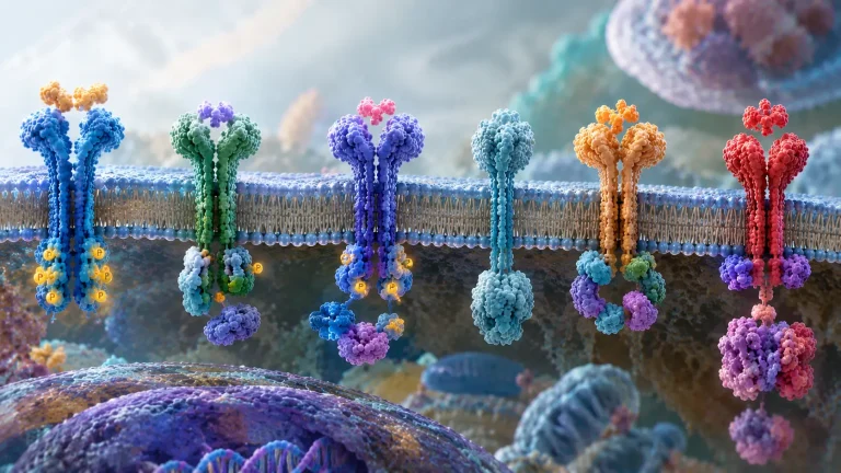
Receptor tyrosine kinases are single-pass transmembrane proteins with three structural parts: an extracellular ligand-binding region, a transmembrane helix, and an intracellular tyrosine kinase domain. The human genome encodes 58 RTKs across roughly 20 subfamilies (Tomuleasa et al., 2024). Most introductory guides teach the four-step activation sequence above and stop there. This article does not stop there.
This article examines four dimensions of receptor tyrosine kinase biology that basic explanations often overlook: catalytic regulation through pseudokinases, structural regulation through preformed dimers, signal selectivity through ligand bias, and therapeutic intervention at different points along the RTK signaling axis. Together, these dimensions reveal that RTK signaling is more diverse and context-dependent than the conventional ligand-induced dimerization model suggests.
The Standard Model: A Useful Starting Point
The Canonical Activation Pathway
Before examining what guides skip, it helps to establish what they teach. The canonical RTK activation pathway follows four steps. First, an extracellular ligand binds the receptor’s ectodomain. Second, ligand binding promotes receptor dimerization or rearrangement into an active dimeric configuration. Third, the intracellular kinase domains trans-autophosphorylate. Fourth, phosphorylated tyrosines recruit adaptor proteins that launch downstream cascades (Lemmon & Schlessinger, 2010).
An important exception to the simple ligand-induced dimerization model is the insulin receptor. It exists as a disulfide-linked α₂β₂ complex before ligand binding. Consequently, insulin activates this preassembled receptor through conformational rearrangement and allosteric regulation, not through assembly of two independent monomers.
Structural Organization and Family Classification
The human genome encodes 58 RTKs, organized into roughly 20 subfamilies based on ectodomain architecture (Tomuleasa et al., 2024). These include the EGFR/ErbB family, insulin receptor family, PDGFR family, VEGFR family, and FGFR family. Each family follows the same three-part structural plan described above.
Downstream Signaling Cascades
The three core downstream pathways are Ras-MAPK, PI3K-Akt, and PLCγ. Ras-MAPK drives proliferation through ERK activation. PI3K-Akt promotes survival by activating Akt and inhibiting apoptosis. PLCγ generates IP3 and DAG, mobilizing calcium and activating protein kinase C.
In addition to these three major pathways, RTKs can engage JAK-STAT and NF-κB signaling networks, depending on receptor subtype, cellular context, and interacting proteins. These pathways do not operate independently. Extensive crosstalk allows RTKs to coordinate proliferation, differentiation, metabolism, inflammation, and survival.
RTK Pseudokinases: Signaling Without Catalytic Activity
The Common Claim and the Evidence
The common claim: RTKs are enzymes. Their name says so. Kinase activity drives signaling.
The evidence: Current literature recognizes eight human receptor tyrosine pseudokinases: HER3 (ErbB3), PTK7, EphB6, EphA10, ROR1, ROR2, RYK, and STYK1 (Park et al., 2026). Five are commonly identified as definitive pseudokinases, while ROR1, ROR2, and RYK are included in broader classifications. Pseudokinase classification concerns conventional phosphotransferase activity, not whether a receptor can participate in phosphorylation-dependent signaling.
How Pseudokinases Function Without Catalysis
Beyond Catalytic Inactivity: Regulatory Roles of Pseudokinases
For years, researchers treated these receptors as inactive remnants. Current evidence shows otherwise. Depending on the receptor, pseudokinases can participate in ligand recognition, associate with catalytically active receptors, regulate receptor complexes, or recruit downstream signaling proteins.
HER3: An Allosteric Activator of HER2
A well-characterized example is HER3 (ErbB3), which exhibits markedly impaired intrinsic kinase activity and therefore depends on catalytically active receptor partners, particularly HER2. However, HER3 is not merely a passive docking platform. Its pseudokinase domain functions as an obligate allosteric activator of the HER2 kinase domain. The original structural evidence for this mechanism emerged from work published in 2009, which demonstrated that the HER3 kinase domain binds ATP yet remains catalytically inactive, and can serve as an activator of the EGFR kinase domain (Jura et al., 2009). Littlefield et al. (2022) later extended this by mapping the ATP-pocket dependence and targetable sites. In the HER2–HER3 heterodimer, the C-lobe of the HER3 kinase domain interfaces with the N-lobe of HER2, stabilizing HER2 in its active conformation. ATP binding to the HER3 pseudokinase domain, despite the absence of catalysis, is critical for this structural stabilization (Littlefield et al., 2022).
Therapeutic Implications of the HER3 ATP-Binding Pocket
An additional implication is that the HER3 ATP-binding pocket may represent a therapeutic target through an unconventional mechanism. Because ATP binding contributes to structural stability rather than supporting phosphotransfer, compounds interacting with this pocket cannot automatically be assumed to inhibit HER3 in the same manner as conventional kinase inhibitors.
ROR and RYK: Pseudokinases in Wnt Signaling
ROR1, ROR2, and RYK participate in Wnt-related signaling, although their ligand-recognition mechanisms differ. ROR1 and ROR2 possess extracellular cysteine-rich domains (CRDs) related to those in Frizzled receptors, whereas RYK contains a Wnt inhibitory factor (WIF) domain. Structural investigations of the Drosophila ROR ortholog Nrk and the RYK ortholog Derailed-2 showed that neither extracellular domain could readily accommodate the acyl chain commonly attached to Wnt proteins. Furthermore, Drosophila Wnt-5 bound these receptors even in the absence of an acyl chain (Shi et al., 2021). These findings suggest that Wnt acylation is not universally required for recognition by ROR and RYK family receptors.
Can Pseudokinases Still Be Targeted by Kinase Inhibitors?
Notably, a counterexample complicates the assumption that pseudokinases cannot be targeted by conventional kinase inhibitors. Sheetz et al. (2020) found that despite the inaccessible ATP-binding pockets of ROR1 and related pseudokinases, screening identified ATP-competitive type-II inhibitors that bind these domains. This demonstrates that even pseudokinases with occluded ATP sites may remain susceptible to small-molecule modulation.
Why This Matters for RTK Biology
Kinase activity is not the only route through which an RTK-family receptor can influence cellular behavior. Scaffolding, allostery, ligand recognition, and heterodimerization can all contribute to signal transmission.
This distinction also matters for drug development. Conventional ATP-competitive inhibitors target catalytic activity, but this approach may not be appropriate for every pseudokinase. Depending on the receptor, alternative strategies may target ligand interactions, protein–protein associations, receptor conformation, or stability.
Preformed RTK Dimers: Activation Without Ligand-Induced Dimerization
Challenging the Ligand-Induced Dimerization Model
The common claim: Ligand binding induces dimerization. No ligand, no dimer, no signal.
The evidence: The insulin receptor family exists as a covalent disulfide-linked dimer before any ligand appears. Ligand binding changes the dimer’s conformation rather than creating it. This is the clearest and most established mammalian example of preformed dimerization among RTKs.
More surprisingly, the Caenorhabditis elegans EGFR homolog LET-23 was shown in 2015 to be constitutively dimeric, yet it responds to its ligand LIN-3 without changing oligomerization state (Freed et al., 2015). This finding first challenged the idea that ligand binding must always initiate receptor dimerization. A 2026 cryo-electron microscopy study extended this work by revealing the structural basis for LET-23 autoinhibition and activation (Zuo et al., 2026). However, the structural mechanism observed in LET-23 should not be assumed to apply universally to mammalian EGFR-family receptors.
Structural Evidence from Insulin Receptors and the LET-23 EGFR Homolog
The insulin receptor exemplifies activation through conformational rearrangement within a preassembled complex. Its α₂β₂ architecture allows insulin binding to trigger changes in receptor organization that activate the intracellular kinase domains without requiring new dimer assembly.
For LET-23, the 2026 cryo-EM study revealed that the membrane-proximal extracellular ends of its preformed dimer are approximately 200 Å apart in the unliganded state. Intermolecular interactions within the cysteine-rich domain II and domain IV hold these “legs” apart, keeping the intracellular kinase domains separated and inactive. Following ligand binding, the membrane-proximal ends move to within 30 Å of each other, bringing the kinase domains into proximity and enabling trans-autophosphorylation (Zuo et al., 2026).
Critically, preformed dimerization modulates ligand sensitivity but is not essential for LET-23 signaling itself. Disrupting the preformed dimer sensitizes the receptor to activation but does not abolish signaling (Zuo et al., 2026). This structural mechanism should not be generalized to every mammalian EGFR-family receptor without further evidence. Notably, the purified extracellular regions of human EGFR family members form only monomers, contrasting with LET-23 (Freed et al., 2015; Zuo et al., 2026).
Cellular studies have described preformed EGFR dimers in living cells (Zuo et al., 2026), and the extent to which membrane context and intracellular interactions stabilize such dimers continues to be investigated. The LET-23 findings should be read as a well-characterized structural example within the broader EGFR family, not as a universal template for all mammalian EGFR-family receptors.
Implications for Receptor Activation Models
These findings show that receptor dimerization and receptor activation are related but distinct events. A receptor may already exist as a dimer while remaining inactive. Ligand binding can then rearrange the receptor into an active configuration.
Consequently, simply measuring receptor dimerization may not provide a reliable indication of RTK activation. Researchers must also consider receptor conformation, kinase-domain association, and phosphorylation status.
Ligand Bias in RTK Signaling: Why Ligands Produce Different Responses
The Concept of Ligand Bias
The common claim: Ligand binds, receptor activates, downstream pathway fires. Different ligands produce the same result.
The evidence: Different ligands can activate the same receptor while producing distinct patterns of phosphorylation. Work on EGFR demonstrated that EGF and TGFα preferentially promote Y1068 phosphorylation over Y1173. In contrast, epiregulin showed no intrinsic preference between these phosphorylation sites in absolute-bias analysis, although it displayed a relative preference for Y1173 when compared with EGF or TGFα (Wirth et al., 2023). These measurements were made in plasma-membrane vesicles, isolating receptor-level phosphorylation from downstream feedback loops. This is a receptor-level result, not a downstream-pathway result.
Earlier work established complementary principles. Freed et al. (2017) showed that EGFR ligands differentially stabilize receptor dimers, with epiregulin and epigen producing less stable dimers than EGF. A 2020 review further consolidated evidence that RTK ligands can produce biased signaling outputs (Karl et al., 2020).
Quantifying Bias in RTK Signaling
Quantifying ligand bias requires more than comparing the magnitude of responses produced by two ligands. Researchers need to compare multiple phosphorylation sites across concentration-response curves, using appropriate reference ligands and analytical methods. Receptor expression, ligand potency, assay conditions, and cellular context must also be considered (Schuck et al., 2026).
Another consideration is the temporal and spatial organization of receptor signaling. Phosphorylation patterns measured at a single time point may not capture differences in signal duration, receptor internalization, or degradation. Comprehensive characterization of RTK ligand bias should ideally combine multiple phosphorylation-site measurements with time-course analyses and downstream functional readouts.
Mutation-Induced Bias in Cancer
Pathogenic mutations can also introduce bias. The EGFR L858R mutation (also designated L834R in an alternative numbering system) changes the relative phosphorylation pattern, favoring Y1173 over Y1068 (Wirth et al., 2023). These findings demonstrate that receptor mutations can influence signaling selectivity, in addition to altering overall receptor activity.
These findings provide a rationale for investigating pathway-selective or mutation-informed inhibitors. However, translating experimentally observed signaling bias into clinically effective therapies remains an important research challenge.
Why Activation Is Not Binary
Activation is not binary. The identity of the ligand, and even the identity of a mutation, can steer the receptor toward different downstream outputs. Nevertheless, experimentally demonstrated ligand bias should not be confused with an established therapeutic strategy. Translating these findings into clinically useful drugs requires evidence that the desired signaling output can be selectively modified without producing unacceptable effects on other essential cellular functions.
Therapeutically Targeting the RTK Signaling Axis Beyond Cancer
Targeting the VEGF Axis in Retinal Disease
The common claim: RTK inhibitors are cancer drugs. Their clinical value lies in oncology.
The evidence: RTK signaling can be targeted at multiple levels, and non-cancer applications extend beyond retinal disease. Pegaptanib provides one example. It is a pegylated RNA aptamer that selectively binds VEGF-A₁₆₅, preventing this ligand from activating VEGF receptors. The FDA approved it in December 2004 for neovascular age-related macular degeneration (U.S. Food and Drug Administration, 2004). Pegaptanib is a VEGF-A₁₆₅ neutralizing agent, not a direct VEGFR tyrosine kinase inhibitor.
Pegaptanib retains historical importance as the first approved agent in this class. Clinically, however, broader anti-VEGF agents have largely superseded it in routine practice.
Axitinib represents a different therapeutic strategy. It is a potent, selective inhibitor of VEGFR-1, VEGFR-2, and VEGFR-3. AXPAXLI, an investigational axitinib-based intravitreal hydrogel developed by Ocular Therapeutix, is designed to provide sustained local inhibition of VEGF receptor signaling. In February 2026, the company reported that its Phase 3 SOL-1 trial met its primary superiority endpoint at Week 36 compared with a single aflibercept injection, with positive findings also reported at Week 52 (Ocular Therapeutix, 2026a). Following a pre-NDA meeting with the FDA in September 2026, the company indicated that it planned to submit an NDA in the fourth quarter of 2026 (Ocular Therapeutix, 2026b). AXPAXLI remains investigational. (These are sponsor-reported results, not independently peer-reviewed findings.)
Kinase Inhibition in Pulmonary Fibrosis
Beyond retinal disease, RTK inhibitors have established roles in other non-cancer conditions. Nintedanib, a multi-kinase inhibitor targeting VEGFR, FGFR, and PDGFR, was approved by the FDA on October 15, 2014 for idiopathic pulmonary fibrosis and remains a standard antifibrotic therapy (U.S. Food and Drug Administration, 2014). The INPULSIS trials demonstrated that nintedanib reduced the annual rate of decline in forced vital capacity compared with placebo (Richeldi et al., 2014).
In October 2025, the FDA approved nerandomilast (Jascayd) for IPF (U.S. Food and Drug Administration, 2025a). Nerandomilast acts through PDE4B inhibition, not through RTK inhibition, and therefore represents a different therapeutic mechanism within the same disease area. It subsequently received approval for progressive pulmonary fibrosis in December 2025 (U.S. Food and Drug Administration, 2025b).
Three Therapeutic Strategies for Targeting RTK Signaling
RTK-directed therapeutic strategies can be grouped into three broad categories, each illustrated here with oncology antibodies, since approved non-oncology RTK antibodies are rarer.
First, ligand neutralization prevents receptor activation by sequestering the extracellular signaling molecule. Pegaptanib, the VEGF-A₁₆₅ aptamer approved for neovascular age-related macular degeneration, exemplifies this approach.
Second, therapeutic antibodies target the extracellular receptor region. Trastuzumab, a humanized monoclonal antibody against HER2, binds the receptor’s extracellular domain. Proposed mechanisms include disruption of HER2–HER3 signaling, receptor downregulation, and recruitment of immune effector cells through antibody-dependent cellular cytotoxicity (Valabrega et al., 2007; Dai et al., 2020). Cetuximab, a chimeric anti-EGFR antibody, competitively inhibits ligand binding to EGFR and promotes receptor internalization (Vincenzi et al., 2010).
Third, small-molecule tyrosine kinase inhibitors act intracellularly by interfering with kinase activity. Nintedanib, a VEGFR/FGFR/PDGFR inhibitor approved for pulmonary fibrosis, exemplifies this strategy in a non-oncology context. The clinical effectiveness and mechanism of each approach depend on the specific ligand, receptor, disease, and therapeutic agent.
These three strategies collectively demonstrate that RTK signaling can be interrupted at the ligand, the receptor, or the kinase domain. RTK signaling controls angiogenesis, metabolism, and development, and its therapeutic reach extends beyond tumor biology into retinal disease, pulmonary fibrosis, and beyond.
Canonical Model vs. Documented Complexity in Receptor Tyrosine Kinases
| Feature | Standard textbook explanation | More complete biological picture |
| Kinase activity | Required for RTK signaling | Pseudokinases can participate in signaling despite lacking conventional catalytic activity; some remain susceptible to ATP-competitive inhibitors |
| Dimerization | Ligand binding induces receptor dimerization | Some receptors exist as preformed dimers (insulin receptor, LET-23), with activation depending on conformational rearrangement |
| Ligand effect | Different ligands produce broadly similar activation | Ligands can generate distinct receptor phosphorylation patterns and signaling outputs |
| Mutation effects | Mutations mainly increase receptor activity | Some mutations can alter signaling preferences |
| Therapeutic scope | RTK targeting is primarily associated with cancer | RTK signaling is targeted by approved therapies beyond cancer: ligand neutralization in retinal disease and kinase inhibition (nintedanib) in pulmonary fibrosis |
What These RTK Signaling Dimensions Mean
Key Lessons for Students
Students should recognize that protein function does not always equal enzymatic activity. Pseudokinases challenge the assumption that a kinase domain must catalyze phosphorylation to matter. Receptor dimerization does not automatically mean receptor activation. Ligand bias demonstrates that receptor activation can produce different signaling outputs.
Implications for Researchers and Experimental Design
Researchers should reconsider experimental design. If a receptor dimerizes before ligand addition, measuring dimerization as an activation readout may mislead. Ligand bias also complicates dose-response interpretation. Demonstrating genuine bias requires comparisons across multiple signaling readouts while accounting for potency, efficacy, receptor expression, and experimental context.
Clinical Relevance for Healthcare Learners
RTK-related signaling is clinically relevant beyond oncology. In retinal disease, anti-VEGF ligand neutralization is established, while sustained-release intravitreal kinase inhibition remains investigational. In pulmonary fibrosis, nintedanib is an approved RTK inhibitor; nerandomilast is also approved for this disease but acts through PDE4B, not an RTK. Understanding the difference between ligand neutralization, receptor-directed antibodies, and intracellular kinase inhibition helps healthcare learners interpret the mechanisms and clinical status of targeted therapies.
Opportunities for Drug Developers
Ligand-bias research may help drug developers identify strategies that selectively modulate particular signaling outputs rather than broadly suppressing receptor activity. Such approaches could eventually improve therapeutic selectivity, although this potential still requires experimental and clinical validation.
Conclusion
Receptor tyrosine kinases are not simple on-off switches. They are sophisticated signaling systems with multiple layers of regulation that introductory explanations often overlook. Pseudokinases participate in signaling without conventional catalytic activity, sometimes through allosteric activation of their partners. Preformed dimers can remain inactive until ligand binding triggers structural rearrangement. Ligand identity and receptor mutations can influence the pattern of downstream signaling. Therapeutic intervention can occur at the level of the ligand, receptor, or intracellular kinase domain.
These complexities do not invalidate the standard model. Instead, they refine it. From an educational perspective, these concepts help students develop a clearer understanding of cellular communication. In research, they reveal experimental limitations while identifying opportunities for further investigation. Within healthcare education, they clarify how targeted therapies achieve their effects through distinct molecular mechanisms.
The dimensions most guides skip are not exceptions to RTK biology. They are an essential part of understanding it.
Frequently Asked Questions
If LET-23 forms dimers before ligand binding, why doesn’t it signal constantly?
In LET-23, the unliganded preformed dimer adopts an autoinhibited conformation. Intermolecular interactions maintain separation between the extracellular juxtamembrane regions at approximately 200 Å, keeping the intracellular kinase domains apart. Ligand binding rearranges the receptor structure, reducing this distance to approximately 30 Å and permitting kinase-domain association and activation .
Can pseudokinases be drug targets?
Yes. Pseudokinases are potential therapeutic targets, although their lack of conventional catalytic activity changes the available strategies. Depending on the receptor, compounds may target ligand interactions, protein–protein associations, receptor conformation, or stability. Importantly, Sheetz et al. (2020) identified ATP-competitive type-II inhibitors for ROR1 despite its inaccessible ATP site, demonstrating that even pseudokinases can be susceptible to small-molecule modulation.
Is ligand bias the same as biased agonism in GPCRs?
The concepts are related but not identical. GPCR biased agonism commonly refers to differential activation of pathways such as G-protein and β-arrestin signaling. In RTKs, ligand bias can involve preferential phosphorylation of distinct tyrosine residues, which recruit different adaptor proteins and influence downstream signaling.
How common are preformed RTK dimers?
Preformed dimers are well-documented for the insulin receptor family and have been structurally characterized for the C. elegans EGFR homolog LET-23. Whether other RTK families form functionally important ligand-independent dimers remains an area of investigation.
Do pseudokinases have any enzymatic function at all?
Most pseudokinases lack conventional protein kinase catalytic activity. However, they can participate in signaling through scaffolding, receptor association, and allosteric mechanisms. HER3 binds ATP with high affinity despite lacking catalytic activity, and this binding serves a structural role in allosteric activation of HER2. By contrast, PTK7, ROR1/2, and RYK do not bind AT.
References
Primary Research Articles
Structural and Mechanistic Studies
- Jura, N., Shan, Y., Cao, X., Shaw, D. E., & Kuriyan, J. (2009). Structural analysis of the catalytically inactive kinase domain of the human EGF receptor 3. Proceedings of the National Academy of Sciences, *106*(51), 21608–21613. https://doi.org/10.1073/pnas.0912101106
Open Access - Freed, D. M., Alvarado, D., & Lemmon, M. A. (2015). Ligand regulation of a constitutively dimeric EGF receptor. Nature Communications, *6*, Article 7380. https://doi.org/10.1038/ncomms8380
Open Access - Freed, D. M., Bessman, N. J., Kiyatkin, A., Salazar-Cavazos, E., Byrne, P. O., Moore, J. O., Valley, C. C., Ferguson, K. M., Leahy, D. J., Lidke, D. S., & Lemmon, M. A. (2017). EGFR ligands differentially stabilize receptor dimers to specify signaling kinetics. Cell, *171*(3), 683–695.e18. https://doi.org/10.1016/j.cell.2017.09.017
Open Access - Sheetz, J. B., Mathea, S., Karvonen, H., Malhotra, K., Chatterjee, D., Niininen, W., Perttilä, R., Preuss, F., Suresh, K., Stayrook, S. E., Tsutsui, Y., Radhakrishnan, R., Ungureanu, D., Knapp, S., & Lemmon, M. A. (2020). Structural insights into pseudokinase domains of receptor tyrosine kinases. Molecular Cell, *79*(3), 390–405.e7. https://doi.org/10.1016/j.molcel.2020.06.018
Open Access - Shi, F., Mendrola, J. M., Sheetz, J. B., Wu, N., Sommer, A., Speer, K. F., Noordermeer, J. N., Kan, Z.-Y., Perry, K., Englander, S. W., Stayrook, S. E., Fradkin, L. G., & Lemmon, M. A. (2021). ROR and RYK extracellular region structures suggest that receptor tyrosine kinases have distinct WNT-recognition modes. Cell Reports, *37*(3), Article 109834. https://doi.org/10.1016/j.celrep.2021.109834
Open Access - Wirth, D., Özdemir, E., & Hristova, K. (2023). Quantification of ligand and mutation-induced bias in EGFR phosphorylation in direct response to ligand binding. Nature Communications, *14*, Article 7579. https://doi.org/10.1038/s41467-023-42926-8
Open Access - Zuo, Y., Schwartz, H. T., Walker, K., Han, L., Sternberg, P. W., & Ferguson, K. M. (2026). Ligand regulation and function of preformed EGFR dimers. Proceedings of the National Academy of Sciences, *123*(30), Article e2602436123. https://doi.org/10.1073/pnas.2602436123
Open Access
HER3 and Pseudokinase Signaling
- Campbell, M. R., Ruiz-Saenz, A., Peterson, E., Agnew, C., Ayaz, P., Garfinkle, S., Littlefield, P., Steri, V., Oeffinger, J., Sampang, M., Shan, Y., Shaw, D. E., Jura, N., & Moasser, M. M. (2022). Targetable HER3 functions driving tumorigenic signaling in HER2-amplified cancers. Cell Reports, 38(5), 110291. https://doi.org/10.1016/j.celrep.2021.110291 (Open Access)
Review Articles and Perspectives
Foundational RTK Reviews
- Lemmon, M. A., & Schlessinger, J. (2010). Cell signaling by receptor tyrosine kinases. Cell, 141(7), 1117–1134. https://doi.org/10.1016/j.cell.2010.06.011 (Open Access)
Pseudokinases
- Park, E. C., Thompson, A. P., Murphy, J. M., Liang, L.-Y., & Lucet, I. S. (2026). The multifaceted roles of receptor tyrosine pseudokinases in cellular signalling. Biochemical Society Transactions, 54(6), 727–743. https://doi.org/10.1042/bst20260770
Ligand Bias
- Karl, K., Paul, M. D., Pasquale, E. B., & Hristova, K. (2020). Ligand bias in receptor tyrosine kinase signaling. Journal of Biological Chemistry, 295(52), 18494–18507. https://doi.org/10.1074/jbc.rev120.015190 (Open Access)
- Schuck, R. J., Roy, T., Bondar, A.-N., Krejci, P., & Hristova, K. (2026). Mechanisms and manifestations of ligand bias in Signaling: Focus On receptor tyrosine kinases. Chemical Reviews, 126(15), 8487–8516. https://doi.org/10.1021/acs.chemrev.6c00023 (Open Access )
Therapeutic Targeting
- Tomuleasa, C., Tigu, A.-B., Munteanu, R., Moldovan, C.-S., Kegyes, D., Onaciu, A., Gulei, D., Ghiaur, G., Einsele, H., & Croce, C. M. (2024). Therapeutic advances of targeting receptor tyrosine kinases in cancer. Signal Transduction and Targeted Therapy, 9(1). https://doi.org/10.1038/s41392-024-01899-w (Open Access)
- Zhang, Y. (2021). The root cause of drug resistance in HER2-positive breast cancer and the therapeutic approaches to overcoming the resistance. Pharmacology & Therapeutics, 218, 107677. https://doi.org/10.1016/j.pharmthera.2020.107677 (Open Access)
- Valabrega, G., Montemurro, F., & Aglietta, M. (2007). Trastuzumab: Mechanism of action, resistance and future perspectives in HER2-overexpressing breast cancer. Annals of Oncology, 18(6), 977–984. https://doi.org/10.1093/annonc/mdl475 (Open Access )
- Vincenzi, B., Zoccoli, A., Pantano, F., Venditti, O., & Galluzzo, S. (2010). CETUXIMAB: From bench to bedside. Current Cancer Drug Targets, 10(1), 80–95. https://doi.org/10.2174/156800910790980241 (Institutional access may be required.)
FDA Approval Letters and Press Releases
- U.S. Food and Drug Administration. (2004). Macugen (pegaptanib sodium) injection approval letter. https://www.accessdata.fda.gov/drugsatfda_docs/appletter/2004/021756ltr.pdf (Open Access)
- U.S. Food and Drug Administration. (2014). OFEV (nintedanib) capsules approval letter (NDA 205832). https://www.accessdata.fda.gov/drugsatfda_docs/appletter/2014/205832Orig1s000ltr.pdf (Open Access)
- U.S. Food and Drug Administration. (2025a, October 7). FDA approves drug to treat idiopathic pulmonary fibrosis [Press release]. https://www.fda.gov/drugs/news-events-human-drugs/fda-approves-drug-treat-idiopathic-pulmonary-fibrosis (Open Access)
- U.S. Food and Drug Administration. (2025b, December 19). FDA approves drug to treat chronic, progressive lung disease [Press release]. https://www.fda.gov/drugs/news-events-human-drugs/fda-approves-drug-treat-chronic-progressive-lung-disease (Open Access)
Industry and Investigational Product Communications
- Ocular Therapeutix, Inc. (2026, February 17). Ocular therapeutixTM reports positive results from landmark SOL-1 phase 3 superiority trial in wet AMD. GlobeNewswire. https://www.globenewswire.com/news-release/2026/02/17/3239086/0/en/ocular-therapeutix-reports-positive-results-from-landmark-sol-1-phase-3-superiority-trial-in-wet-amd.html (Open Access)
- Ocular Therapeutix, Inc. (2026, September 15). Ocular therapeutixTM announces positive pre-nda meeting with U.S. FDA for AXPAXLITM in wet AMD. GlobeNewswire. https://www.globenewswire.com/news-release/2026/09/15/3361910/0/en/ocular-therapeutix-announces-positive-pre-nda-meeting-with-u-s-fda-for-axpaxli-in-wet-amd.html (Open Access)