
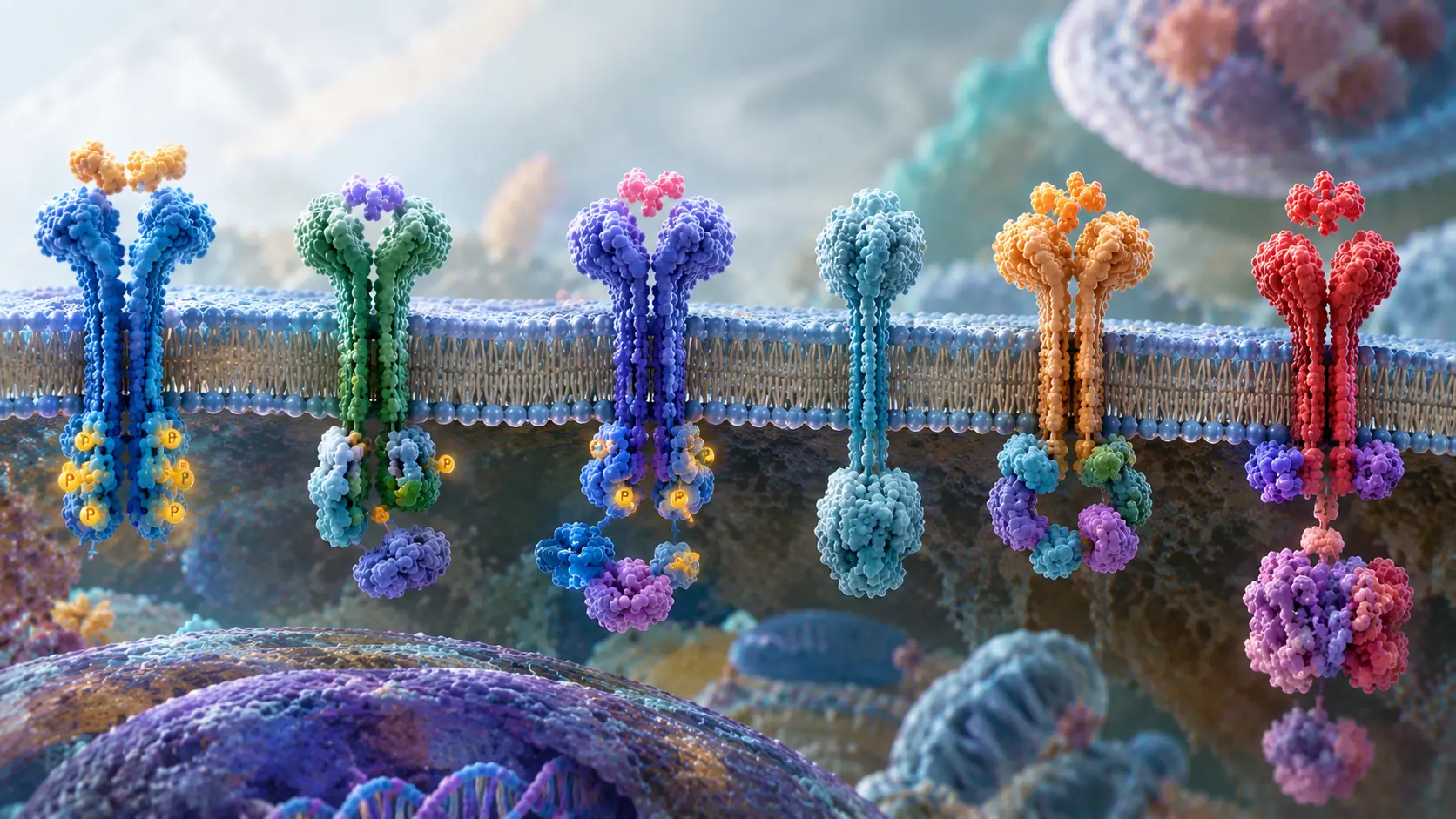
Enzyme-Linked Receptors: Unlocking Cell Signaling and Disease
Introduction: How Do Enzyme-Linked Receptors Control Cell Behavior?
What if a single protein on a cell’s surface could decide whether that cell grows, divides, or dies? Enzyme linked receptors do exactly that. They sit at the boundary between the outside world and the cell’s interior. When a chemical message arrives, they convert it into action.
Enzyme-linked receptors act as molecular switches. While some possess intrinsic enzymatic activity, others recruit enzymes to initiate downstream signaling. In either case, they convert extracellular signals into a cascade of biochemical reactions within the cell (Alberts et al., 2015). Consequently, these receptors are fundamental to understanding molecular biology and cellular communication. Students encounter them early in their molecular biology studies, whereas researchers investigating cancer and other diseases continue to explore their roles. Importantly, when these receptors malfunction, the consequences can be severe. Indeed, dysregulated signaling through enzyme-linked receptors is implicated in several human diseases, including cancer, immune disorders, and metabolic diseases (Zhang & Li, 2023).
This guide walks through their architecture, activation steps, major families, and disease connections. Short analogies and real examples keep the science approachable. By the end, you will understand why these receptors remain prime drug targets and how they shape human health.
What Are Enzyme-Linked Receptors?
Enzyme-linked receptors, also known as catalytic receptors, are single-pass transmembrane proteins characterized by three key structural components. First, their extracellular domain binds specific ligands, such as growth factors or hormones. Next, a single transmembrane helix anchors the receptor within the cell membrane. Finally, the intracellular region either contains an intrinsic catalytic domain or associates closely with an enzyme to initiate downstream signaling (Alberts et al., 2015).
The defining feature of enzyme-linked receptors is their direct coupling to intracellular enzymatic activity. Once ligand binding occurs, the receptor undergoes a conformational change that activates its associated enzyme or intrinsic catalytic domain. In contrast to G protein-coupled receptors, which transmit signals through intermediate G proteins, enzyme-linked receptors activate downstream signaling pathways more directly. Consequently, their signaling responses are often slower in onset but more sustained (Raj et al., 2026).
Think of the receptor as a molecular switch. The ligand flips the switch. The enzyme does the work. The result is a phosphorylation cascade that alters gene expression, metabolism, or cell shape.
Structure of Enzyme-Linked Receptors
Most enzyme-linked receptors share a relatively simple structural organization, although their individual components may vary. In particular, the extracellular region exhibits considerable diversity and determines the specificity of ligand recognition. For example, some receptors contain immunoglobulin-like domains, whereas others possess cysteine-rich regions.
In contrast, the transmembrane segment typically consists of a short, hydrophobic helix that anchors the receptor within the cell membrane. Finally, the cytoplasmic tail contains either the catalytic domain itself or specific docking sites for associated enzymes, thereby facilitating intracellular signal transmission (Alberts et al., 2015).
Receptor tyrosine kinases (RTKs) constitute the largest group of enzyme-linked receptors. These receptors contain an intracellular tyrosine kinase domain that transfers phosphate groups from ATP to specific tyrosine residues on target proteins. Similarly, receptor serine/threonine kinases catalyse the phosphorylation of serine or threonine residues. In contrast, guanylyl cyclase receptors function differently by catalysing the conversion of GTP into cyclic guanosine monophosphate (cGMP), an important intracellular second messenger. Tyrosine-kinase-associated receptors lack their own kinase but recruit Janus kinases or Src-family kinases (Raj et al., 2026).
Dimerization is a common feature of enzyme-linked receptor activation. In many cases, receptors exist as individual monomers until a ligand binds. Subsequently, ligand binding promotes the association of two receptor molecules, bringing their intracellular domains into close proximity and enabling their mutual activation. However, not all receptors require ligand-induced dimerization. For example, insulin receptors exist as pre-formed, disulfide-linked dimers that undergo conformational rearrangements upon insulin binding, thereby initiating intracellular signaling (Alberts et al., 2015).
How Enzyme-Linked Receptors Transmit Signals
Receptor activation generally follows a well-organized sequence of molecular events. Initially, the ligand binds to the extracellular domain of the receptor. Subsequently, receptor molecules undergo dimerization or oligomerization, bringing their intracellular catalytic domains into close proximity. As a result, these catalytic domains become activated. In the case of receptor tyrosine kinases (RTKs), activation involves autophosphorylation of specific tyrosine residues. Importantly, the resulting phosphotyrosine residues serve as docking platforms for intracellular signaling proteins containing Src homology 2 (SH2) or phosphotyrosine-binding (PTB) domains, thereby facilitating the recruitment of downstream signaling components (Zhang & Li, 2023).
Adapter proteins such as Grb2 recruit guanine nucleotide exchange factors that turn on Ras. Ras then launches the MAPK cascade that drives proliferation. Phosphoinositide 3-kinase binds other phosphotyrosines and generates PIP3, activating Akt and promoting survival. Parallel pathways can modulate calcium, cytoskeletal dynamics, or transcription factors (Zhang & Li, 2023).
A short analogy helps. Imagine two people standing apart holding switches. When a ball, the ligand, pulls them together, their switches click on. A chain of lights, phosphorylation events, eventually reaches the control room, the nucleus.
Termination is equally important. Protein tyrosine phosphatases remove the phosphates. Receptor internalization and degradation clear the signal. Feedback loops keep the system balanced (von Zastrow & Sorkin, 2021).
Major Types of Enzyme-Linked Receptors
Five principal classes dominate textbooks and research. A sixth category, receptor tyrosine phosphatases, is sometimes included but requires careful qualification. For a quick side-by-side view of all five classes, including their catalytic activities and representative members, see Table 1 below.
| Table 1. The Five Main Classes of Enzyme-Linked Receptors | |||
| Class | Catalytic Activity | Key Examples | Primary Functions |
| Receptor tyrosine kinases | Intrinsic tyrosine kinase | EGFR, insulin receptor, VEGFR, Trk | Growth, metabolism, angiogenesis |
| Tyrosine-kinase-associated | Associated JAK or Src kinases | Cytokine receptors (EPO, IL receptors) | Immunity, hematopoiesis |
| Receptor serine/threonine kinases | Intrinsic Ser/Thr kinase | TGF-β receptors, BMP receptors | Development, differentiation, fibrosis |
| Receptor guanylyl cyclases | Intrinsic guanylyl cyclase | ANP receptors (NPR1, NPR2) | Blood pressure, fluid balance |
| Histidine-kinase-associated | Histidine phosphorylation | Bacterial sensor kinases | Environmental sensing (microbial) |
This table 1 summarizes the major families of enzyme linked receptors, their enzymatic activities, representative members, and physiological roles. Data compiled from standard cell biology sources (Alberts et al., 2015; Raj et al., 2026).
Receptor Tyrosine Kinases
Receptor tyrosine kinases (RTKs) constitute the largest and most extensively studied family of enzyme-linked receptors. In humans, this family comprises 58 distinct receptors classified into approximately 20 subfamilies (Raj et al., 2026). Among the well-characterized members are the epidermal growth factor receptor (EGFR), insulin receptor (IR), vascular endothelial growth factor receptors (VEGFRs), and neurotrophin receptors (NTRs). Collectively, these receptors regulate a wide range of cellular processes, including cell growth, metabolism, angiogenesis, survival, and neuronal development.
These receptors control growth, differentiation, metabolism, and angiogenesis. Their dysfunction is central to many cancers. The erbB family was first linked to cancer when researchers discovered that the avian erythroblastosis virus encoded an altered form of the human epidermal growth factor (EGF) receptor. Consequently, this finding established an important connection between erbB family genes and cancer development. (Zhang & Li, 2023). Drugs such as gefitinib and erlotinib block the kinase domain and have transformed treatment of certain lung cancers (Zhang & Li, 2023).
Tyrosine-Kinase-Associated Receptors
Cytokine receptors lack intrinsic kinase activity. After ligand binding, they recruit Janus kinases, which phosphorylate the receptor and STATs. The JAK-STAT pathway rapidly alters gene expression and is central to immune responses.
Humans have four JAKs: JAK1, JAK2, JAK3, and TYK2. Each interacts with specific cytokine receptors and recruits corresponding STATs (Djidjik et al., 2025). Loss-of-function mutations weaken cytokine signaling, thereby compromising immune function and leading to immunodeficiency. In contrast, gain-of-function mutations cause excessive or dysregulated signaling, which can contribute to autoimmune diseases and myeloproliferative syndromes. (Djidjik et al., 2025). Negative regulators such as SOCS proteins and SHP-1/SHP-2 phosphatases keep the pathway in check.
Receptor Serine/Threonine Kinases
The transforming growth factor-beta (TGF-β) receptor family serves as a classic example of enzyme-linked receptors. Upon ligand binding, type I and type II receptors come together to form a functional receptor complex. Next, the type II receptor phosphorylates and activates the type I receptor. In turn, the activated type I receptor phosphorylates Smad proteins, which subsequently translocate to the nucleus and regulate gene transcription (Massagué & Sheppard, 2023).
Beyond their roles in cellular signaling, these receptors play essential roles in embryonic patterning, wound healing, and immune tolerance. However, their biological effects are highly context-dependent and may vary considerably depending on the cellular environment and disease state. For example, transforming growth factor-beta (TGF-β) can suppress tumor development in certain contexts; conversely, it may promote tumor progression and metastasis in others. Because of these seemingly opposing effects, TGF-β has earned the nickname “Jekyll and Hyde growth factor” (Massagué & Sheppard, 2023).
Receptor Guanylyl Cyclases
These receptors generate the second messenger cGMP. The atrial natriuretic peptide receptors are classic examples. Binding of ANP activates the intracellular cyclase domain, raises cGMP, and promotes vasodilation and natriuresis.
A critical detail sets this family apart. Receptor guanylyl cyclase activity depends on phosphorylation. For GC-A and GC-B, phosphorylation of four to seven conserved serine or threonine residues maintains activity, while dephosphorylation inactivates the enzyme. This regulatory mechanism controls cardiac hypertrophy, long bone growth, and bone density (Potter, 2024).
Histidine-Kinase-Associated Systems
Similarly, bacteria employ two-component signaling systems to detect and respond to changes in their environment. In these systems, a sensor histidine kinase first undergoes autophosphorylation on a conserved histidine residue. Subsequently, it transfers the phosphate group to a specific aspartate residue on its corresponding response regulator, thereby activating the regulator and triggering an appropriate cellular response. These systems matter in microbial biotechnology but differ from mammalian enzyme linked receptors (Alberts et al., 2015).
Downstream Pathways and Cellular Outcomes
Once activated, enzyme-linked receptors initiate several conserved intracellular signaling pathways. Among these, the Ras–MAPK pathway promotes the transcription of genes required for cell-cycle progression and proliferation. Similarly, the PI3K–Akt–mTOR signaling axis supports protein synthesis, glucose uptake, and cell survival. Another important pathway involves phospholipase C-gamma (PLC-γ), which generates inositol 1,4,5-trisphosphate (IP3) and diacylglycerol (DAG), thereby mobilizing intracellular calcium and activating protein kinase C (PKC) (Zhang & Li, 2023).
In contrast, transforming growth factor-beta (TGF-β) receptors primarily signal through the Smad pathway. Following receptor activation, phosphorylated Smad proteins associate with other Smad family members and transcriptional co-regulators to regulate specific sets of target genes. Importantly, these signaling pathways rarely operate independently. Instead, extensive cross-talk with G protein-coupled receptor (GPCR) and Wnt signaling pathways adds further layers of regulation. Consequently, the cellular response depends heavily on the biological context. For instance, the same epidermal growth factor receptor (EGFR) signal may promote cell proliferation in one cell type while inducing differentiation in another. The major signaling pathways, their key components, and their disease relevance are compared in greater detail in Table 2.
| Table 2. Signaling Pathways and Disease Relevance | |||
| Pathway | Key Components | Main Functions | Disease Relevance |
| RAS-MAPK | RAS, RAF, MEK, ERK | Proliferation, differentiation | Cancer, developmental disorders |
| PI3K-AKT | PI3K, PIP3, AKT, PTEN | Survival, metabolism, growth | Cancer, metabolic dysregulation |
| JAK-STAT | JAK1/2/3, TYK2, STATs | Immune and hormonal responses | Immune dysfunction, inflammation |
| TGF-β-SMAD | TβRII, TβRI, SMAD2/3, SMAD4 | Differentiation, matrix regulation | Fibrosis, cancer |
| cGMP signaling | Guanylyl cyclase, cGMP | Cardiovascular, renal, skeletal | Cardiovascular disorders |
These pathways represent major signaling routes downstream of enzyme linked receptors. Their effects overlap, and the final response depends on receptor activity, cellular context, and interactions with other pathways (Zhang & Li, 2023; Massagué & Sheppard, 2023; Potter, 2024).
Real-World Examples of Enzyme-Linked Receptors in Action
Consider insulin signaling as an example. When blood glucose levels rise, insulin binds to its receptor, triggering receptor autophosphorylation and the recruitment of insulin receptor substrate (IRS) proteins. Subsequently, downstream PI3K–Akt signaling promotes the translocation of GLUT4 transporters to the cell membrane, thereby facilitating glucose uptake. Consequently, defects in this signaling pathway can contribute to the development of type 2 diabetes (Alberts et al., 2015).
EGFR offers another story. In developing tissues, EGF binding stimulates epithelial growth. In many cancers, the receptor is overexpressed or mutated, producing constant growth signals.
VEGF receptors guide new blood-vessel formation. Tumors exploit this pathway to secure their own blood supply. Anti-VEGF antibodies and tyrosine-kinase inhibitors remain mainstays of oncology (Zhang & Li, 2023).
A brief mini-story illustrates the point. A researcher studying wound healing notices that fibroblasts migrate faster when PDGF is present. Blocking the PDGF receptor tyrosine kinase slows migration and reduces scarring. The same receptor, when overactive in the wrong context, can promote fibrosis.
Enzyme-Linked Receptors Versus Other Receptor Classes
G protein-coupled receptors (GPCRs) span the cell membrane seven times and transmit signals through heterotrimeric G proteins. Consequently, their responses are often rapid and involve second messengers such as cyclic adenosine monophosphate (cAMP) and inositol 1,4,5-trisphosphate (IP3). In contrast, enzyme-linked receptors typically cross the membrane only once and mediate intracellular signaling through enzymatic activity, particularly protein phosphorylation. Meanwhile, ligand-gated ion channels respond to ligand binding by opening ion-conducting pores, often within milliseconds. By comparison, nuclear receptors bind lipid-soluble ligands within the cell and subsequently regulate gene transcription, producing responses that may develop over hours (Alberts et al., 2015).
Enzyme linked receptors therefore occupy a middle ground. They are slower than ion channels yet faster and more direct than many nuclear-receptor effects. Therefore, their outputs frequently change gene expression and long-term cell fate. For a structured comparison of enzyme linked receptors with GPCRs and ion channels, see Table 3 below.
| Table 3. Comparison of Cell-Surface Receptor Classes | |||
| Feature | Enzyme-Linked Receptors | GPCRs | Ion Channels |
| Transmembrane passes | Single | Seven | Multiple |
| Initial mechanism | Enzyme activation | G protein activation | Ion pore opening |
| Response speed | Slower, longer-lasting | Rapid to intermediate | Milliseconds |
| Example | EGFR | β-adrenergic receptor | Nicotinic acetylcholine receptor |
This table 3 highlights key differences between enzyme linked receptors and other major cell-surface receptor families (Alberts et al., 2015).
Physiological Importance and Disease Links
During embryonic development, enzyme-linked receptors play essential roles in tissue patterning and organ formation. For example, fibroblast growth factor (FGF) and epidermal growth factor (EGF) receptors contribute to processes such as limb development and neural induction. Following development, these receptors continue to perform important functions in adult tissues by maintaining tissue homeostasis, regulating metabolism, and coordinating immune responses (Raj et al., 2026).
However, disruption of these signaling pathways can have serious pathological consequences. For instance, activating mutations or gene amplifications involving receptor tyrosine kinases (RTKs) can drive the development and progression of several cancers. Similarly, loss or impairment of TGF-β signaling may remove important cellular growth restraints, thereby contributing to tumor development. In addition, mutations affecting natriuretic peptide receptors can disrupt cardiovascular regulation, particularly blood pressure control. Consequently, advances in understanding receptor-mediated signaling pathways have enabled the development of targeted therapies for various human diseases. These drugs include inhibitors that act on intracellular kinase domains as well as therapeutic agents that target extracellular receptor regions (Zhang & Li, 2023).
Recent reviews highlight that more than fifty small-molecule receptor tyrosine kinase (RTK) inhibitors, along with several monoclonal antibodies, have entered clinical use (Zhang & Li, 2023). Nevertheless, therapeutic resistance remains a major challenge, often limiting treatment effectiveness. To address this limitation, researchers are exploring combination therapies and next-generation protein degraders, which continue to expand the range of available targeted treatment strategies.
Practical Insights for Students and Researchers
When you read a paper that mentions an RTK, ask three questions. Which ligand activates it? Does the receptor dimerize? Which downstream pathway is dominant in that cell type? Sketching the pathway on paper solidifies the knowledge. Laboratory work often begins with Western blots for phosphotyrosine or with specific kinase inhibitors to test necessity (von Zastrow & Sorkin, 2021).
For content creators and educators, analogies help. Compare receptor dimerization to two puzzle pieces clicking together. Compare autophosphorylation to a light switch that stays on until a phosphatase turns it off.
Conclusion: Why Enzyme-Linked Receptors Matter
Enzyme linked receptors stand among the most versatile and medically important cell-surface sensors. They convert extracellular cues into intracellular enzymatic activity through dimerization, phosphorylation, and carefully orchestrated cascades. From insulin controlling blood sugar to EGFR driving tissue growth and cancer, these receptors shape both health and disease.
Mastering their structure, the five main classes, and the core signaling routes equips students, clinicians, and researchers with a powerful conceptual toolkit (Alberts et al., 2015; Raj et al., 2026). The next time you encounter a growth-factor pathway or a targeted cancer therapy, remember that enzyme linked receptors sit at the center of the story.
Keep exploring primary literature. Test the pathways in model systems. Share the insights so the next generation can build on them.
Frequently Asked Questions About Enzyme-Linked Receptors
What are enzyme-linked receptors?
Enzyme-linked receptors are cell-surface proteins that transmit signals into cells by activating their own enzymatic domains or associating with intracellular enzymes. Through this signaling process, they regulate essential cellular activities, including growth, differentiation, metabolism, and immune responses.
What are the main types of enzyme-linked receptors?
The main types include receptor tyrosine kinases, tyrosine-kinase-associated receptors, receptor serine/threonine kinases, and receptor guanylyl cyclases. Although these receptor classes share a common role in signal transmission, each uses a distinct enzymatic mechanism to regulate cellular activities.
How do enzyme-linked receptors work?
When a ligand binds to an enzyme-linked receptor, it activates the receptor or an associated enzyme. Subsequently, this activation initiates intracellular signaling pathways, such as RAS–MAPK, PI3K–AKT, and JAK–STAT. As a result, these pathways regulate gene expression, metabolism, cell growth, and survival.
What is the difference between enzyme-linked receptors and G protein-coupled receptors?
Enzyme-linked receptors typically cross the cell membrane once and activate intrinsic or associated enzymes. In contrast,G protein-coupled receptors (GPCRs) cross the membrane seven times and transmit signals through heterotrimeric G proteins. Despite these structural and functional differences, both receptor classes regulate essential cellular responses through distinct signaling mechanisms.
How are enzyme-linked receptors involved in cancer?
Abnormal receptor activation, caused by mutations, gene amplification, or excessive receptor expression, can disrupt normal cell-growth control. For example, altered epidermal growth factor receptor (EGFR) signaling can promote uncontrolled cell proliferation and contribute to cancer development. Consequently, several targeted cancer therapies inhibit receptor tyrosine kinases or their downstream signaling pathways.
References
- Alberts B, Johnson A, Lewis J, et al. Molecular Biology of the Cell. 4th edition. New York: Garland Science; 2002. Signaling through Enzyme-Linked Cell-Surface Receptors. Available from: https://www.ncbi.nlm.nih.gov/books/NBK26822/ (Open access via NCBI Bookshelf)
- Djidjik R, Lamara Mahammed L, Berkani LM, Allam I, Benmoussa AH, Gharnaout M and Belaid B (2025) JAK/STAT in human diseases: a common axis in immunodeficiencies and hematological disorders. Front. Immunol. 16:1669688. https://doi.org/10.3389/fimmu.2025.1669688 (Open access)
- Massagué, J., & Sheppard, D. (2023). TGF-β signaling in health and disease. Cell, 186(19), 4007–4037. https://doi.org/10.1016/j.cell.2023.07.036 (Open access via PubMed Central)
- Lincoln R Potter, Phosphorylation-Dependent Regulation of Guanylyl Cyclase (GC)-A and Other Membrane GC Receptors, Endocrine Reviews, Volume 45, Issue 5, October 2024, Pages 755–771, https://doi.org/10.1210/endrev/bnae015 (Open access)
- Raj, A., Abu-Sailik, F., Jawabri, A. A., Ahmad, B. M., Mohamed, F. E., et al. (2026). Receptor tyrosine kinase families: A central hub of cellular signaling and involvement in human health and disease. Human Genomics. https://doi.org/10.1186/s40246-026-01042-6 (Open access)
- von Zastrow, M., & Sorkin, A. (2021). Mechanisms for regulating and organizing receptor signaling by endocytosis. Annual Review of Biochemistry, 90, 709–737. https://doi.org/10.1146/annurev-biochem-081820-092427 (Open access via PubMed Central)
- Zhang, N., & Li, Y. (2023). Receptor tyrosine kinases: Biological functions and anticancer targeted therapy. MedComm, 4(6), Article e446. https://doi.org/10.1002/mco2.446 (Open access)
About Author
Dr. Niamat Khan, PhD, is an Assistant Professor of Biotechnology and Genetic Engineering, molecular geneticist, and genomics researcher with over 19 years of teaching and research experience. His interests include human genetics, inherited disorders, hereditary cancers, bioinformatics, and AI in genomic medicine.
Through The Scholar Post, he simplifies complex scientific concepts for students, researchers, educators, and science enthusiasts through research-based educational articles.
Explore his academic background and research: Dr. Niamat Khan, PhD – Author Profile